 Movie
Movie Controller

[English] 日本語
 Yorodumi
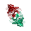
- PDB-1n71: Crystal structure of aminoglycoside 6'-acetyltransferase type Ii ... -
+ Open data

- Basic information
| Entry | Database: PDB / ID: 1n71 | ||||||
|---|---|---|---|---|---|---|---|
| Title | Crystal structure of aminoglycoside 6'-acetyltransferase type Ii in complex with coenzyme A | ||||||
 Components Components | aac(6')-Ii | ||||||
 Keywords Keywords | TRANSFERASE / AMINOGLYCOSIDE 6'-N-ACETYLTRANSFERASE / ANTIBIOTIC RESISTANCE / COENZYME A | ||||||
| Function / homology |  Function and homology information Function and homology informationacyltransferase activity, transferring groups other than amino-acyl groups Similarity search - Function | ||||||
| Biological species |  Enterococcus faecium (bacteria) Enterococcus faecium (bacteria) | ||||||
| Method |  X-RAY DIFFRACTION / SYNCHROTRON / MOLECULAR REPLACEMENT / Resolution: 1.8 Å X-RAY DIFFRACTION / SYNCHROTRON / MOLECULAR REPLACEMENT / Resolution: 1.8 Å | ||||||
 Authors Authors | Burk, D.L. / Ghuman, N. / Wybenga-Groot, L.E. / Berghuis, A.M. | ||||||
 Citation Citation | Journal: Protein Sci. / Year: 2003 Title: X-ray structure of the AAC(6')-Ii antibiotic resistance enzyme at 1.8 A resolution; examination of oligomeric arrangements in GNAT superfamily members Authors: Burk, D.L. / Ghuman, N. / Wybenga-Groot, L.E. / Berghuis, A.M. #1: Journal: Structure / Year: 1999Title: Crystal structure of an aminoglycoside 6'-N-acetyltransferase: defining the GCN5-related N-acetyltransferase superfamily fold Authors: Wybenga-Groot, L.E. / Draker, K. / Wright, G.D. / Berghuis, A.M. | ||||||
| History |
| ||||||
| Remark 999 | SEQUENCE The author states that there is an error in the sequence database. |
- Structure visualization
| Structure viewer | Molecule: MolmilJmol/JSmol |
|---|
- Downloads & links
-Download
| PDBx/mmCIF format | 1n71.cif.gz | 168.5 KB | Display | PDBx/mmCIF format |
|---|---|---|---|---|
| PDB format | pdb1n71.ent.gz | 135.1 KB | Display | PDB format |
| PDBx/mmJSON format | 1n71.json.gz | Tree view | PDBx/mmJSON format | |
| Others |  Other downloads Other downloads |
-Validation report
| Arichive directory | https://data.pdbj.org/pub/pdb/validation_reports/n7/1n71ftp://data.pdbj.org/pub/pdb/validation_reports/n7/1n71 | HTTPS FTP |
|---|
-Related structure data
| Related structure data |  1b87S S: Starting model for refinement |
|---|---|
| Similar structure data |


-Links
 PDBj
PDBj


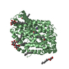
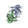
- Assembly
| Deposited unit | 
| ||||||||
|---|---|---|---|---|---|---|---|---|---|
| 1 | 
| ||||||||
| 2 | 
| ||||||||
| 3 | 
| ||||||||
| Unit cell |
| ||||||||
| Details | Asymmetric unit contains two biological units (dimers) |
-Components
| #1: Protein | Mass: 20514.988 Da / Num. of mol.: 4 Source method: isolated from a genetically manipulated source Source: (gene. exp.) Enterococcus faecium (bacteria) / Plasmid: pPLaac-1 / Production host: #2: Chemical | ChemComp-COA /   Mass: 767.534 Da / Num. of mol.: 4 / Source method: obtained synthetically / Formula: C21H36N7O16P3S Mass: 767.534 Da / Num. of mol.: 4 / Source method: obtained synthetically / Formula: C21H36N7O16P3S#3: Chemical | ChemComp-SO4 /   Mass: 96.063 Da / Num. of mol.: 5 / Source method: obtained synthetically / Formula: SO4 Mass: 96.063 Da / Num. of mol.: 5 / Source method: obtained synthetically / Formula: SO4#4: Water | ChemComp-HOH / |  Mass: 18.015 Da / Num. of mol.: 504 / Source method: isolated from a natural source / Formula: H2O Mass: 18.015 Da / Num. of mol.: 504 / Source method: isolated from a natural source / Formula: H2O |
|---|
-Experimental details
-Experiment
| Experiment | Method: X-RAY DIFFRACTION / Number of used crystals: 1 |
|---|
- Sample preparation
| Crystal | Density Matthews: 2.02 Å3/Da / Density % sol: 38.54 % | ||||||||||||||||||||
|---|---|---|---|---|---|---|---|---|---|---|---|---|---|---|---|---|---|---|---|---|---|
| Crystal grow | Temperature: 295 K / Method: vapor diffusion, hanging drop / pH: 7.5 Details: ammonium sulfate, pH 7.5, VAPOR DIFFUSION, HANGING DROP, temperature 295K | ||||||||||||||||||||
| Crystal grow | *PLUS Temperature: 22 ℃ | ||||||||||||||||||||
| Components of the solutions | *PLUS
|
-Data collection
| Diffraction | Mean temperature: 110 K |
|---|---|
| Diffraction source | Source: SYNCHROTRON / Site: NSLS  / Beamline: X8C / Wavelength: 1.0722 Å / Beamline: X8C / Wavelength: 1.0722 Å |
| Detector | Type: ADSC QUANTUM 4 / Detector: CCD / Date: Apr 29, 2000 |
| Radiation | Protocol: SINGLE WAVELENGTH / Monochromatic (M) / Laue (L): M / Scattering type: x-ray |
| Radiation wavelength | Wavelength: 1.0722 Å / Relative weight: 1 |
| Reflection | Resolution: 1.8→27.97 Å / Num. all: 64971 / Num. obs: 64971 / % possible obs: 94.4 % / Observed criterion σ(F): 0 / Observed criterion σ(I): 0 / Redundancy: 5.6 % / Biso Wilson estimate: 19.2 Å2 / Rmerge(I) obs: 0.08 / Net I/σ(I): 17.6 |
| Reflection shell | Resolution: 1.8→1.86 Å / Redundancy: 1.4 % / Rmerge(I) obs: 0.44 / Mean I/σ(I) obs: 3.1 |
| Reflection | *PLUS Highest resolution: 1.8 Å / Lowest resolution: 100 Å / Rmerge(I) obs: 0.08 |
| Reflection shell | *PLUS % possible obs: 67.6 % / Redundancy: 3.1 % / Num. unique obs: 4590 / Mean I/σ(I) obs: 1.4 |
- Processing
| Software |
| |||||||||||||||||||||||||
|---|---|---|---|---|---|---|---|---|---|---|---|---|---|---|---|---|---|---|---|---|---|---|---|---|---|---|
| Refinement | Method to determine structure: MOLECULAR REPLACEMENT Starting model: pdb entry 1B87 with cofactor and water removed Resolution: 1.8→27.97 Å / Rfactor Rfree error: 0.003 / Isotropic thermal model: RESTRAINED / Cross valid method: THROUGHOUT / σ(F): 0 / σ(I): 0 / Stereochemistry target values: Engh & Huber
| |||||||||||||||||||||||||
| Solvent computation | Solvent model: FLAT MODEL / Bsol: 45.5792 Å2 / ksol: 0.359296 e/Å3 | |||||||||||||||||||||||||
| Displacement parameters | Biso mean: 26.1 Å2
| |||||||||||||||||||||||||
| Refine analyze | Luzzati coordinate error free: 0.26 Å / Luzzati sigma a free: 0.2 Å | |||||||||||||||||||||||||
| Refinement step | Cycle: LAST / Resolution: 1.8→27.97 Å
| |||||||||||||||||||||||||
| Refine LS restraints |
| |||||||||||||||||||||||||
| LS refinement shell | Resolution: 1.8→1.91 Å / Rfactor Rfree error: 0.011 / Total num. of bins used: 6
| |||||||||||||||||||||||||
| Xplor file |
| |||||||||||||||||||||||||
| Refinement | *PLUS % reflection Rfree: 10 % / Rfactor Rfree: 0.241 / Rfactor Rwork: 0.203 | |||||||||||||||||||||||||
| Solvent computation | *PLUS | |||||||||||||||||||||||||
| Displacement parameters | *PLUS | |||||||||||||||||||||||||
| Refine LS restraints | *PLUS
|
