 Movie
Movie Controller

[English] 日本語
 Yorodumi
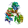
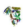
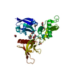
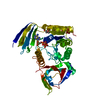
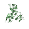
- PDB-2a2d: X-ray structure of human n-acetyl galactosamine kinase complexed ... -
+ Open data

- Basic information
| Entry | Database: PDB / ID: 2a2d | |||||||||
|---|---|---|---|---|---|---|---|---|---|---|
| Title | X-ray structure of human n-acetyl galactosamine kinase complexed with Mn-AMPPNP and n-acetyl glactosamine | |||||||||
 Components Components | N-acetylgalactosamine kinase | |||||||||
 Keywords Keywords | TRANSFERASE / kinase / galactokinase | |||||||||
| Function / homology |  Function and homology information Function and homology informationN-acetylgalactosamine kinase / N-acetylgalactosamine kinase activity / galactokinase activity / galactose metabolic process / carbohydrate metabolic process / ATP binding / cytosol Similarity search - Function | |||||||||
| Biological species |  Homo sapiens (human) Homo sapiens (human) | |||||||||
| Method |  X-RAY DIFFRACTION / MOLECULAR REPLACEMENT / Resolution: 2.2 Å X-RAY DIFFRACTION / MOLECULAR REPLACEMENT / Resolution: 2.2 Å | |||||||||
 Authors Authors | Thoden, J.B. / Holden, H.M. | |||||||||
 Citation Citation | Journal: J.Biol.Chem. / Year: 2005 Title: The molecular architecture of human N-acetylgalactosamine kinase. Authors: Thoden, J.B. / Holden, H.M. | |||||||||
| History |
|
- Structure visualization
| Structure viewer | Molecule: MolmilJmol/JSmol |
|---|
- Downloads & links
-Download
| PDBx/mmCIF format | 2a2d.cif.gz | 111.2 KB | Display | PDBx/mmCIF format |
|---|---|---|---|---|
| PDB format | pdb2a2d.ent.gz | 82 KB | Display | PDB format |
| PDBx/mmJSON format | 2a2d.json.gz | Tree view | PDBx/mmJSON format | |
| Others |  Other downloads Other downloads |
-Validation report
| Arichive directory | https://data.pdbj.org/pub/pdb/validation_reports/a2/2a2dftp://data.pdbj.org/pub/pdb/validation_reports/a2/2a2d | HTTPS FTP |
|---|
-Related structure data
| Related structure data |  2a2cSC S: Starting model for refinement C: citing same article ( |
|---|---|
| Similar structure data |


-Links
 PDBj
PDBj


- Assembly
| Deposited unit | 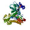
| ||||||||
|---|---|---|---|---|---|---|---|---|---|
| 1 |
| ||||||||
| Unit cell |
|
-Components
| #1: Protein | Mass: 52788.641 Da / Num. of mol.: 1 Source method: isolated from a genetically manipulated source Source: (gene. exp.) Homo sapiens (human) / Gene: GALK2, GK2 / Plasmid: pET28 / Production host:  References: UniProt: Q01415, Transferases; Transferring phosphorus-containing groups; Phosphotransferases with an alcohol group as acceptor |
|---|---|
| #2: Sugar | ChemComp-A2G /   Type: D-saccharide, alpha linking / Mass: 221.208 Da / Num. of mol.: 1 Type: D-saccharide, alpha linking / Mass: 221.208 Da / Num. of mol.: 1Source method: isolated from a genetically manipulated source Formula: C8H15NO6 |
| #3: Chemical | ChemComp-MN /   Mass: 54.938 Da / Num. of mol.: 1 / Source method: obtained synthetically / Formula: Mn Mass: 54.938 Da / Num. of mol.: 1 / Source method: obtained synthetically / Formula: Mn |
| #4: Chemical | ChemComp-ANP /   Mass: 506.196 Da / Num. of mol.: 1 / Source method: obtained synthetically / Formula: C10H17N6O12P3 / Comment: AMP-PNP, energy-carrying molecule analogue*YM Mass: 506.196 Da / Num. of mol.: 1 / Source method: obtained synthetically / Formula: C10H17N6O12P3 / Comment: AMP-PNP, energy-carrying molecule analogue*YM |
| #5: Water | ChemComp-HOH /  Mass: 18.015 Da / Num. of mol.: 256 / Source method: isolated from a natural source / Formula: H2O Mass: 18.015 Da / Num. of mol.: 256 / Source method: isolated from a natural source / Formula: H2O |
-Experimental details
-Experiment
| Experiment | Method: X-RAY DIFFRACTION / Number of used crystals: 1 |
|---|
- Sample preparation
| Crystal | Density Matthews: 2.5 Å3/Da / Density % sol: 50 % |
|---|---|
| Crystal grow | Temperature: 298 K / Method: vapor diffusion, hanging drop / pH: 6 Details: PEG-3400, Mn-AMPPNP, NaCl, MES, n-acetyl galactosamine, pH 6, VAPOR DIFFUSION, HANGING DROP, temperature 298K |
-Data collection
| Diffraction | Mean temperature: 100 K |
|---|---|
| Diffraction source | Source: ROTATING ANODE / Type: RIGAKU RU200 / Wavelength: 1.5418 / Wavelength: 1.5418 Å |
| Detector | Type: Bruker Platinum 135 / Detector: CCD / Date: Apr 29, 2005 / Details: montel optics |
| Radiation | Monochromator: montel optics / Protocol: SINGLE WAVELENGTH / Monochromatic (M) / Laue (L): M / Scattering type: x-ray |
| Radiation wavelength | Wavelength: 1.5418 Å / Relative weight: 1 |
| Reflection | Resolution: 2.2→30 Å / Num. all: 26919 / Num. obs: 26919 / % possible obs: 98.9 % / Observed criterion σ(F): 0 / Observed criterion σ(I): 0 / Redundancy: 8.7 % / Rsym value: 0.081 / Net I/σ(I): 15.9 |
| Reflection shell | Resolution: 2.2→2.3 Å / Redundancy: 3 % / Mean I/σ(I) obs: 3.5 / Num. unique all: 3322 / Rsym value: 0.354 / % possible all: 98.4 |
- Processing
| Software |
| |||||||||||||||||||||||||
|---|---|---|---|---|---|---|---|---|---|---|---|---|---|---|---|---|---|---|---|---|---|---|---|---|---|---|
| Refinement | Method to determine structure: MOLECULAR REPLACEMENT Starting model: PDB entry 2a2c Resolution: 2.2→30 Å / Cross valid method: THROUGHOUT / σ(F): 0 / Stereochemistry target values: Engh & Huber
| |||||||||||||||||||||||||
| Refinement step | Cycle: LAST / Resolution: 2.2→30 Å
| |||||||||||||||||||||||||
| Refine LS restraints |
|
