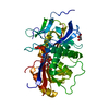
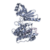
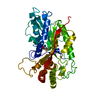
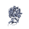
Isomerases; Isomerases altering macromolecular conformation; Enzymes altering nucleic acid conformation / DNA topoisomerase type I (single strand cut, ATP-independent) activity / DNA 5'-3' helicase / 5'-3' DNA helicase activity / ATP hydrolysis activity / DNA binding / ATP binding / metal ion binding / cytoplasm Similarity search - Function
DNA helicase, TraI type / Conjugative transfer relaxase protein TraI / TraI, 2B/2B-like domain / TraI, N-terminal subdomain / : / DNA helicase TraI, C-terminal / single-stranded DNA binding TraI N-terminal subdomain / DNA relaxase TraI 2B/2B-like domain / TraI helicase-associated ssDNA bd, N-terminal / Conjugative relaxase, N-terminal ...DNA helicase, TraI type / Conjugative transfer relaxase protein TraI / TraI, 2B/2B-like domain / TraI, N-terminal subdomain / : / DNA helicase TraI, C-terminal / single-stranded DNA binding TraI N-terminal subdomain / DNA relaxase TraI 2B/2B-like domain / TraI helicase-associated ssDNA bd, N-terminal / Conjugative relaxase, N-terminal / TrwC relaxase / TrwC relaxase / AAA domain / P-loop containing nucleoside triphosphate hydrolase Similarity search - Domain/homology
In the structure databanks used in Yorodumi, some data are registered as the other names, "COVID-19 virus" and "2019-nCoV". Here are the details of the virus and the list of structure data.
Jan 31, 2019. EMDB accession codes are about to change! (news from PDBe EMDB page)
EMDB accession codes are about to change! (news from PDBe EMDB page)
The allocation of 4 digits for EMDB accession codes will soon come to an end. Whilst these codes will remain in use, new EMDB accession codes will include an additional digit and will expand incrementally as the available range of codes is exhausted. The current 4-digit format prefixed with “EMD-” (i.e. EMD-XXXX) will advance to a 5-digit format (i.e. EMD-XXXXX), and so on. It is currently estimated that the 4-digit codes will be depleted around Spring 2019, at which point the 5-digit format will come into force.
The EM Navigator/Yorodumi systems omit the EMD- prefix.
Related info.:Q: What is EMD? / ID/Accession-code notation in Yorodumi/EM Navigator
Yorodumi is a browser for structure data from EMDB, PDB, SASBDB, etc.
This page is also the successor to EM Navigator detail page, and also detail information page/front-end page for Omokage search.
The word "yorodu" (or yorozu) is an old Japanese word meaning "ten thousand". "mi" (miru) is to see.
Related info.:EMDB / PDB / SASBDB / Comparison of 3 databanks / Yorodumi Search / Aug 31, 2016. New EM Navigator & Yorodumi / Yorodumi Papers / Jmol/JSmol / Function and homology information / Changes in new EM Navigator and Yorodumi
 Movie
Movie Controller

 Yorodumi
 Open data
 Basic information
 Components
 Keywords
 Function and homology information
 X-RAY DIFFRACTION /
  Authors
 Citation
 Structure visualization
 Downloads & links
 Other downloads


 PDBj
PDBj


 Assembly

 Mass: 24.305 Da / Num. of mol.: 1 / Source method: obtained synthetically / Formula: Mg
 Mass: 69.085 Da / Num. of mol.: 1 / Source method: obtained synthetically / Formula: C3H5N2
 Mass: 18.015 Da / Num. of mol.: 67 / Source method: isolated from a natural source / Formula: H2O
 Sample preparation
 Processing