 Movie
Movie Controller

[English] 日本語
 Yorodumi
- PDB-207d: SOLUTION STRUCTURE OF MITHRAMYCIN DIMERS BOUND TO PARTIALLY OVERL... -
+ Open data

- Basic information
| Entry | Database: PDB / ID: 207d | |||||||||||||||||||||||
|---|---|---|---|---|---|---|---|---|---|---|---|---|---|---|---|---|---|---|---|---|---|---|---|---|
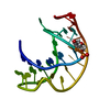
| Title | SOLUTION STRUCTURE OF MITHRAMYCIN DIMERS BOUND TO PARTIALLY OVERLAPPING SITES ON DNA | |||||||||||||||||||||||
 Components Components | DNA (5'-D(* Keywords KeywordsDNA / DOUBLE HELIX / MITHRAMYCIN | Function / homology | Chem-CRH / DNA |  Function and homology information Function and homology informationBiological species | synthetic construct (others) | Method | SOLUTION NMR / molecular dynamics |  Authors AuthorsSastry, M. / Fiala, R. / Patel, D.J. |  CitationJournal: J.Mol.Biol. / Year: 1995 CitationJournal: J.Mol.Biol. / Year: 1995Title: Solution structure of mithramycin dimers bound to partially overlapping sites on DNA. Authors: Sastry, M. / Fiala, R. / Patel, D.J. History |
|
- Structure visualization
| Structure viewer | Molecule: MolmilJmol/JSmol |
|---|
- Downloads & links
-Download
| PDBx/mmCIF format | 207d.cif.gz | 218 KB | Display | PDBx/mmCIF format |
|---|---|---|---|---|
| PDB format | pdb207d.ent.gz | 183.7 KB | Display | PDB format |
| PDBx/mmJSON format | 207d.json.gz | Tree view | PDBx/mmJSON format | |
| Others |  Other downloads Other downloads |
-Validation report
| Arichive directory | https://data.pdbj.org/pub/pdb/validation_reports/07/207dftp://data.pdbj.org/pub/pdb/validation_reports/07/207d | HTTPS FTP |
|---|
-Related structure data
| Similar structure data |
|---|
-Links
 PDBj
PDBj

- Assembly
| Deposited unit | 
| |||||||||
|---|---|---|---|---|---|---|---|---|---|---|
| 1 |
| |||||||||
| NMR ensembles |
|
-Components
| #1: DNA chain | Mass: 3044.017 Da / Num. of mol.: 2 / Source method: obtained synthetically / Details: CHEMICALLY SYNTHESIZED / Source: (synth.) synthetic construct (others) #2: Polysaccharide | beta-D-Olivopyranose-(1-3)-beta-D-Olivopyranose Source method: isolated from a genetically manipulated source #3: Polysaccharide | 2,6-dideoxy-3-C-methyl-beta-D-ribo-hexopyranose-(1-3)-2,6-dideoxy-beta-D-galactopyranose-(1-3)-beta- ...2,6-dideoxy-3-C-methyl-beta-D-ribo-hexopyranose-(1-3)-2,6-dideoxy-beta-D-galactopyranose-(1-3)-beta-D-Olivopyranose #4: Chemical |   Mass: 24.305 Da / Num. of mol.: 2 / Source method: obtained synthetically / Formula: Mg Mass: 24.305 Da / Num. of mol.: 2 / Source method: obtained synthetically / Formula: Mg#5: Chemical | ChemComp-CRH /   Mass: 388.411 Da / Num. of mol.: 4 / Source method: obtained synthetically / Formula: C21H24O7 Mass: 388.411 Da / Num. of mol.: 4 / Source method: obtained synthetically / Formula: C21H24O7 |
|---|
-Experimental details
-Experiment
| Experiment | Method: SOLUTION NMR |
|---|---|
| NMR details | Text: EACH MODEL CONTAINS FOUR MOLECULES OF MITHRAMYCIN (EACH WITH FORMULA C52 H75 O24, CHARGE -1) AND TWO MG++ CATIONS THAT COORDINATE TO TWO MITHRAMYCIN MOLECULES. IN THIS ENTRY MITHRAMYCIN IS ...Text: EACH MODEL CONTAINS FOUR MOLECULES OF MITHRAMYCIN (EACH WITH FORMULA C52 H75 O24, CHARGE -1) AND TWO MG++ CATIONS THAT COORDINATE TO TWO MITHRAMYCIN MOLECULES. IN THIS ENTRY MITHRAMYCIN IS PRESENTED AS HET GROUPS DDA-DDA-CHR- TSC. THE TSC RESIDUE IS COMPRISED OF THREE SACCHARIDE UNITS (DDA-DDL-MDA). THE HYDROPHILIC SIDE CHAIN AND THE A-B DISACCHARIDE OF MITHRAMYCIN MOLECULES ALONG WITH THE TERMINAL BASE PAIR IN THE DNA DUPLEX ARE NOT WELL DEFINED. |
- Sample preparation
| Crystal grow | *PLUS Method: other / Details: NMR |
|---|
- Processing
| Software |
| ||||||||
|---|---|---|---|---|---|---|---|---|---|
| NMR software | Name:  X-PLOR / Developer: BRUNGER / Classification: refinement X-PLOR / Developer: BRUNGER / Classification: refinement | ||||||||
| Refinement | Method: molecular dynamics / Software ordinal: 1 Details: TWO STARTING STRUCTURES WERE OBTAINED BY MANUALLY DOCKING MITHRAMYCIN ON A FORM AND B FORM DNA. THESE WERE SUBSEQUENTLY REFINED BY DISTANCE-RESTRAINED MOLECULAR DYNAMICS USING A SET OF INTER- ...Details: TWO STARTING STRUCTURES WERE OBTAINED BY MANUALLY DOCKING MITHRAMYCIN ON A FORM AND B FORM DNA. THESE WERE SUBSEQUENTLY REFINED BY DISTANCE-RESTRAINED MOLECULAR DYNAMICS USING A SET OF INTER-PROTON DISTANCES DERIVED FROM NMR DATA (40, 80, 120, 160, 250 MS NOESY EXPERIMENTS) AND DELTA DIHEDRAL ANGLES DERIVED FROM SIMULATION OF COSY CROSS PEAK PATTERNS. THE EIGHT DISTANCE RESTRAINED STRUCTURES WERE OBTAINED BY TAKING THE AVERAGE COORDINATES OF THE LAST 2.0 PS OF THE DYNAMICS DURING DISTANCE RESTRAINED DYNAMICS AND MINIMIZED. THE RMS DEVIATIONS FROM IDEAL GEOMETRY FOR THE EIGHT FINAL STRUCTURES ARE: BOND (ANG): MDL1 MDL2 MDL3 MDL4 MDL5 MDL6 MDL7 MDL8 0.009 0.009 0.009 0.008 0.009 0.009 0.009 0.009 ANGLE (DEG): MDL1 MDL2 MDL3 MDL4 MDL5 MDL6 MDL7 MDL8 2.627 2.630 2.631 2.636 2.621 2.638 2.664 2.652 IMPROPER(DEG): MDL1 MDL2 MDL3 MDL4 MDL5 MDL6 MDL7 MDL8 0.866 0.850 0.808 0.874 0.841 0.803 0.881 0.808 | ||||||||
| NMR ensemble | Conformer selection criteria: all calculated structures submitted Conformers calculated total number: 8 / Conformers submitted total number: 8 |
