 Movie
Movie Controller

[English] 日本語
 Yorodumi
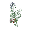
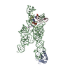
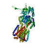
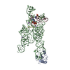
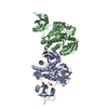
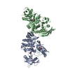
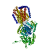
- PDB-1zzn: Crystal structure of a group I intron/two exon complex that inclu... -
+ Open data

- Basic information
| Entry | Database: PDB / ID: 1zzn | ||||||
|---|---|---|---|---|---|---|---|
| Title | Crystal structure of a group I intron/two exon complex that includes all catalytic metal ion ligands. | ||||||
 Components Components |
| ||||||
 Keywords Keywords | STRUCTURAL PROTEIN/RNA / RNA structure / ribozyme / self-splicing intron / Azoarcus / two-metal-ion mechanism / STRUCTURAL PROTEIN-RNA COMPLEX | ||||||
| Function / homology |  Function and homology information Function and homology informationU1 snRNP binding / U1 snRNP / U1 snRNA binding / U4/U6 x U5 tri-snRNP complex / mRNA Splicing - Major Pathway / spliceosomal complex / mRNA splicing, via spliceosome / DNA binding / RNA binding / nucleoplasm ...U1 snRNP binding / U1 snRNP / U1 snRNA binding / U4/U6 x U5 tri-snRNP complex / mRNA Splicing - Major Pathway / spliceosomal complex / mRNA splicing, via spliceosome / DNA binding / RNA binding / nucleoplasm / identical protein binding / nucleus Similarity search - Function | ||||||
| Biological species |  Homo sapiens (human) Homo sapiens (human) | ||||||
| Method |  X-RAY DIFFRACTION / SYNCHROTRON / FOURIER SYNTHESIS / Resolution: 3.37 Å X-RAY DIFFRACTION / SYNCHROTRON / FOURIER SYNTHESIS / Resolution: 3.37 Å | ||||||
 Authors Authors | Stahley, M.R. / Strobel, S.A. | ||||||
 Citation Citation | Journal: Science / Year: 2005 Title: Structural evidence for a two-metal-ion mechanism of group I intron splicing. Authors: Stahley, M.R. / Strobel, S.A. #1: Journal: Nature / Year: 2004Title: Crystal Structure of a Self-Splicing Group I Intron with Both Exons Authors: Adams, P.L. / Stahley, M.R. / Kosek, A.B. / Wang, J. / Strobel, S.A. #2: Journal: RNA / Year: 2004 Title: Crystal Structure of a group I intron splicing intermediate Authors: Adams, P.L. / Stahley, M.R. / Gill, M.L. / Kosek, A.B. / Wang, J. / Strobel, S.A. #3: Journal: Cell(Cambridge,Mass.) / Year: 1981 Title: In vitro splicing of the ribosomal RNA precursor of tetrahymena: involvement of a guanosine nucleotide in the excision of intervening sequence Authors: Cech, T.R. / Zaug, A.J. / Grabowaski, P.J. #4: Journal: Nature / Year: 1992Title: Self-splicing intron in tRNA genes of widely divergent bacteria Authors: Reinhold-Hurek, B. / Shub, D.A. | ||||||
| History |
| ||||||
| Remark 999 | SEQUENCE NUCLEOTIDES 1001B TO 1014B BELONG TO AN ENGINEERED U1A LOOP AND ARE INSERTED BETWEEN GUA- ...SEQUENCE NUCLEOTIDES 1001B TO 1014B BELONG TO AN ENGINEERED U1A LOOP AND ARE INSERTED BETWEEN GUA-107B AND CYT-112B. THERE ARE NO NUCLEOTIDES BETWEEN GUA-1B AND GUA-5B DUE TO A CLONING DESIGN. CHAINS B AND C ARE TWO PARTS OF THE ORIGINAL ONE INTRON SEQUENCE BECAUSE OF AN EXPERIMENTAL DESIGN. THE FIRST THREE RESIDUES IN U1A PROTEIN WERE MISSING IN THE STRUCTURE. |
- Structure visualization
| Structure viewer | Molecule: MolmilJmol/JSmol |
|---|
- Downloads & links
-Download
| PDBx/mmCIF format | 1zzn.cif.gz | 164.1 KB | Display | PDBx/mmCIF format |
|---|---|---|---|---|
| PDB format | pdb1zzn.ent.gz | 119.1 KB | Display | PDB format |
| PDBx/mmJSON format | 1zzn.json.gz | Tree view | PDBx/mmJSON format | |
| Others |  Other downloads Other downloads |
-Validation report
| Arichive directory | https://data.pdbj.org/pub/pdb/validation_reports/zz/1zznftp://data.pdbj.org/pub/pdb/validation_reports/zz/1zzn | HTTPS FTP |
|---|
-Related structure data
| Related structure data |  1u6bS S: Starting model for refinement |
|---|---|
| Similar structure data |


-Links
 PDBj
PDBj


- Assembly
| Deposited unit | 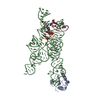
| ||||||||
|---|---|---|---|---|---|---|---|---|---|
| 1 |
| ||||||||
| Unit cell |
|
-Components
-RNA chain , 3 types, 3 molecules BCD
| #1: RNA chain | Mass: 64057.023 Da / Num. of mol.: 1 / Source method: obtained synthetically Details: RNA WAS IS TRANSCRIBED BY T7 RNA POLYMERASE. SEQUENCE FROM AZOARCUS GROUP I INTRON |
|---|---|
| #2: RNA chain | Mass: 7045.354 Da / Num. of mol.: 1 / Source method: obtained synthetically Details: solid phase sythesis. Sequence from Azoarcus intron and exon sequence. |
| #3: RNA chain | Mass: 909.623 Da / Num. of mol.: 1 / Source method: obtained synthetically Details: Solid phase synthesis. Sequence from Azoarcus group I intron 5'-exon sequence. |
-Protein , 1 types, 1 molecules A
| #4: Protein | Mass: 11340.315 Da / Num. of mol.: 1 / Fragment: RRM 1 / Mutation: Y31H, Q36R Source method: isolated from a genetically manipulated source Source: (gene. exp.) Homo sapiens (human) / Gene: SNRPA / Plasmid: pet11 / Species (production host): Escherichia coli / Production host:  |
|---|
-Non-polymers , 3 types, 60 molecules 


| #5: Chemical | ChemComp-MG /  Mass: 24.305 Da / Num. of mol.: 5 / Source method: obtained synthetically / Formula: Mg Mass: 24.305 Da / Num. of mol.: 5 / Source method: obtained synthetically / Formula: Mg#6: Chemical | ChemComp-K / |  Mass: 39.098 Da / Num. of mol.: 1 / Source method: obtained synthetically / Formula: K Mass: 39.098 Da / Num. of mol.: 1 / Source method: obtained synthetically / Formula: K#7: Water | ChemComp-HOH / | Mass: 18.015 Da / Num. of mol.: 54 / Source method: isolated from a natural source / Formula: H2O |
|---|
-Experimental details
-Experiment
| Experiment | Method: X-RAY DIFFRACTION / Number of used crystals: 1 |
|---|
- Sample preparation
| Crystal | Density Matthews: 4.6 Å3/Da / Density % sol: 69.8 % |
|---|---|
| Crystal grow | Temperature: 298 K / Method: vapor diffusion, hanging drop / pH: 6.8 Details: MPD, sodium cacodylate, magnesium acetate, potassium chloride, cobalt heximine, pH 6.8, VAPOR DIFFUSION, HANGING DROP, temperature 298K |
-Data collection
| Diffraction | Mean temperature: 100 K |
|---|---|
| Diffraction source | Source: SYNCHROTRON / Site: NSLS  / Beamline: X25 / Wavelength: 1.1 Å / Beamline: X25 / Wavelength: 1.1 Å |
| Detector | Type: ADSC QUANTUM 315 / Detector: CCD / Date: Jun 29, 2004 |
| Radiation | Monochromator: crystal / Protocol: SINGLE WAVELENGTH / Monochromatic (M) / Laue (L): M / Scattering type: x-ray |
| Radiation wavelength | Wavelength: 1.1 Å / Relative weight: 1 |
| Reflection | Resolution: 3.37→50 Å / Num. all: 20658 / Num. obs: 20658 / % possible obs: 95.9 % / Observed criterion σ(F): 2 / Observed criterion σ(I): 2 / Redundancy: 5.5 % / Rmerge(I) obs: 0.072 / Net I/σ(I): 18.2 |
| Reflection shell | Resolution: 3.37→3.49 Å / Redundancy: 5.6 % / Mean I/σ(I) obs: 1.1 / % possible all: 71.1 |
- Processing
| Software |
| ||||||||||||||||||||||||||||||||||||||||||||||||||||||||||||||||||||||||||||||||
|---|---|---|---|---|---|---|---|---|---|---|---|---|---|---|---|---|---|---|---|---|---|---|---|---|---|---|---|---|---|---|---|---|---|---|---|---|---|---|---|---|---|---|---|---|---|---|---|---|---|---|---|---|---|---|---|---|---|---|---|---|---|---|---|---|---|---|---|---|---|---|---|---|---|---|---|---|---|---|---|---|---|
| Refinement | Method to determine structure: FOURIER SYNTHESIS Starting model: PDB entry 1U6B Resolution: 3.37→33.04 Å / Rfactor Rfree error: 0.011 / Data cutoff high absF: 198973.33 / Data cutoff low absF: 0 / Isotropic thermal model: RESTRAINED / Cross valid method: THROUGHOUT / σ(F): 0 / Stereochemistry target values: CNS Details: Experimental phases from 1U6B structure were used followed by maximum likelihood refinement. Octahedral parameters for Mg2+ coordination were used to refine M1 and M2 in the final round of refinement only
| ||||||||||||||||||||||||||||||||||||||||||||||||||||||||||||||||||||||||||||||||
| Solvent computation | Solvent model: FLAT MODEL / Bsol: 10 Å2 / ksol: 0.0527796 e/Å3 | ||||||||||||||||||||||||||||||||||||||||||||||||||||||||||||||||||||||||||||||||
| Displacement parameters | Biso mean: 100.7 Å2
| ||||||||||||||||||||||||||||||||||||||||||||||||||||||||||||||||||||||||||||||||
| Refine analyze |
| ||||||||||||||||||||||||||||||||||||||||||||||||||||||||||||||||||||||||||||||||
| Refinement step | Cycle: LAST / Resolution: 3.37→33.04 Å
| ||||||||||||||||||||||||||||||||||||||||||||||||||||||||||||||||||||||||||||||||
| Refine LS restraints |
| ||||||||||||||||||||||||||||||||||||||||||||||||||||||||||||||||||||||||||||||||
| LS refinement shell | Resolution: 3.37→3.58 Å / Rfactor Rfree error: 0.039 / Total num. of bins used: 6
| ||||||||||||||||||||||||||||||||||||||||||||||||||||||||||||||||||||||||||||||||
| Xplor file |
|
