 Movie
Movie Controller

[English] 日本語
 Yorodumi
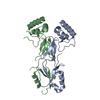
- PDB-1zxz: X-ray structure of peptide deformylase from Arabidopsis thaliana ... -
+ Open data

- Basic information
| Entry | Database: PDB / ID: 1zxz | ||||||
|---|---|---|---|---|---|---|---|
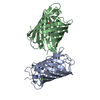
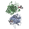
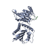
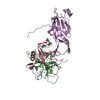
| Title | X-ray structure of peptide deformylase from Arabidopsis thaliana (AtPDF1A); crystals grown in PEG-5000 MME as precipitant | ||||||
 Components Components | Peptide deformylase, mitochondrial | ||||||
 Keywords Keywords | HYDROLASE / peptide deformylase / PDF1A / eukaryote / higher plant / Arabidopsis thaliana / zinc ion | ||||||
| Function / homology |  Function and homology information Function and homology informationplant-type cell wall / peptide deformylase / peptide deformylase activity / chloroplast stroma / chloroplast / protein maturation / translation / mitochondrion / metal ion binding Similarity search - Function | ||||||
| Biological species |  | ||||||
| Method |  X-RAY DIFFRACTION / SYNCHROTRON / MOLECULAR REPLACEMENT / Resolution: 2.8 Å X-RAY DIFFRACTION / SYNCHROTRON / MOLECULAR REPLACEMENT / Resolution: 2.8 Å | ||||||
 Authors Authors | Fieulaine, S. / Juillan-Binard, C. / Serero, A. / Dardel, F. / Giglione, C. / Meinnel, T. / Ferrer, J.-L. | ||||||
 Citation Citation | Journal: J.Biol.Chem. / Year: 2005 Title: The crystal structure of mitochondrial (Type 1A) peptide deformylase provides clear guidelines for the design of inhibitors specific for the bacterial forms Authors: Fieulaine, S. / Juillan-Binard, C. / Serero, A. / Dardel, F. / Giglione, C. / Meinnel, T. / Ferrer, J.-L. | ||||||
| History |
|
- Structure visualization
| Structure viewer | Molecule: MolmilJmol/JSmol |
|---|
- Downloads & links
-Download
| PDBx/mmCIF format | 1zxz.cif.gz | 91 KB | Display | PDBx/mmCIF format |
|---|---|---|---|---|
| PDB format | pdb1zxz.ent.gz | 68.2 KB | Display | PDB format |
| PDBx/mmJSON format | 1zxz.json.gz | Tree view | PDBx/mmJSON format | |
| Others |  Other downloads Other downloads |
-Validation report
| Arichive directory | https://data.pdbj.org/pub/pdb/validation_reports/zx/1zxzftp://data.pdbj.org/pub/pdb/validation_reports/zx/1zxz | HTTPS FTP |
|---|
-Related structure data
| Related structure data |  1zy0C  1zy1C  1y6hS C: citing same article ( S: Starting model for refinement |
|---|---|
| Similar structure data |


-Links
 PDBj
PDBj- Assembly
| Deposited unit | 
| ||||||||||
|---|---|---|---|---|---|---|---|---|---|---|---|
| 1 |
| ||||||||||
| Unit cell |
| ||||||||||
| Details | Dimer in the asymmetric unit, but it is unclear whether it is a biological dimer or not |
-Components
| #1: Protein | Mass: 22310.689 Da / Num. of mol.: 2 / Fragment: mature protein Source method: isolated from a genetically manipulated source Source: (gene. exp.)  #2: Chemical |   Mass: 65.409 Da / Num. of mol.: 2 / Source method: obtained synthetically / Formula: Zn Mass: 65.409 Da / Num. of mol.: 2 / Source method: obtained synthetically / Formula: Zn#3: Water | ChemComp-HOH / |  Mass: 18.015 Da / Num. of mol.: 210 / Source method: isolated from a natural source / Formula: H2O Mass: 18.015 Da / Num. of mol.: 210 / Source method: isolated from a natural source / Formula: H2O |
|---|
-Experimental details
-Experiment
| Experiment | Method: X-RAY DIFFRACTION / Number of used crystals: 1 |
|---|
- Sample preparation
| Crystal | Density Matthews: 2.5 Å3/Da / Density % sol: 50.2 % |
|---|---|
| Crystal grow | Temperature: 291 K / Method: vapor diffusion, hanging drop / pH: 5.5 Details: Crystals grown in PEG-5000 MME as precipitant, pH 5.5, VAPOR DIFFUSION, HANGING DROP, temperature 291K |
-Data collection
| Diffraction | Mean temperature: 100 K |
|---|---|
| Diffraction source | Source: SYNCHROTRON / Site: ESRF  / Beamline: BM30A / Wavelength: 0.979 Å / Beamline: BM30A / Wavelength: 0.979 Å |
| Detector | Type: MARRESEARCH 176mm / Detector: CCD / Date: Jan 30, 2005 |
| Radiation | Monochromator: 2 silicon 111 / Protocol: SINGLE WAVELENGTH / Monochromatic (M) / Laue (L): M / Scattering type: x-ray |
| Radiation wavelength | Wavelength: 0.979 Å / Relative weight: 1 |
| Reflection | Resolution: 2.8→50 Å / Num. all: 10698 / Num. obs: 10552 / % possible obs: 99 % / Observed criterion σ(I): 1 / Rmerge(I) obs: 0.105 / Rsym value: 0.105 / Net I/σ(I): 13.2 |
- Processing
| Software |
| |||||||||||||||||||||||||
|---|---|---|---|---|---|---|---|---|---|---|---|---|---|---|---|---|---|---|---|---|---|---|---|---|---|---|
| Refinement | Method to determine structure: MOLECULAR REPLACEMENT Starting model: 1Y6H Resolution: 2.8→30 Å / Cross valid method: THROUGHOUT / σ(F): 2 / Stereochemistry target values: Engh & Huber
| |||||||||||||||||||||||||
| Refine analyze |
| |||||||||||||||||||||||||
| Refinement step | Cycle: LAST / Resolution: 2.8→30 Å
| |||||||||||||||||||||||||
| Refine LS restraints |
|
