 Movie
Movie Controller


+ Open data

- Basic information
| Entry | Database: PDB / ID: 1zx4 | ||||||
|---|---|---|---|---|---|---|---|
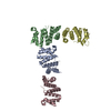
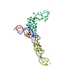
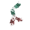
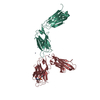
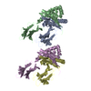
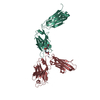
| Title | Structure of ParB bound to DNA | ||||||
 Components Components |
| ||||||
 Keywords Keywords | CELL CYCLE / partition / P1 / plasmid | ||||||
| Function / homology |  Function and homology information Function and homology information | ||||||
| Biological species |  Enterobacteria phage P1 (virus) Enterobacteria phage P1 (virus) | ||||||
| Method |  X-RAY DIFFRACTION / SYNCHROTRON / MAD / Resolution: 2.98 Å X-RAY DIFFRACTION / SYNCHROTRON / MAD / Resolution: 2.98 Å | ||||||
 Authors Authors | Schumacher, M.A. / Funnell, B.E. | ||||||
 Citation Citation | Journal: Nature / Year: 2005 Title: Structures of ParB bound to DNA reveal mechanism of partition complex formation. Authors: Schumacher, M.A. / Funnell, B.E. | ||||||
| History |
|
- Structure visualization
| Structure viewer | Molecule: MolmilJmol/JSmol |
|---|
- Downloads & links
-Download
| PDBx/mmCIF format | 1zx4.cif.gz | 114.2 KB | Display | PDBx/mmCIF format |
|---|---|---|---|---|
| PDB format | pdb1zx4.ent.gz | 85.2 KB | Display | PDB format |
| PDBx/mmJSON format | 1zx4.json.gz | Tree view | PDBx/mmJSON format | |
| Others |  Other downloads Other downloads |
-Validation report
| Summary document | 1zx4_validation.pdf.gz | 473.7 KB | Display | wwPDB validaton report |
|---|---|---|---|---|
| Full document | 1zx4_full_validation.pdf.gz | 510.7 KB | Display | |
| Data in XML | 1zx4_validation.xml.gz | 22.9 KB | Display | |
| Data in CIF | 1zx4_validation.cif.gz | 30.2 KB | Display | |
| Arichive directory | https://data.pdbj.org/pub/pdb/validation_reports/zx/1zx4ftp://data.pdbj.org/pub/pdb/validation_reports/zx/1zx4 | HTTPS FTP |
-Related structure data
| Similar structure data |
|---|
-Links
 PDBj
PDBj


- Assembly
| Deposited unit | 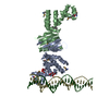
| ||||||||||
|---|---|---|---|---|---|---|---|---|---|---|---|
| 1 |
| ||||||||||
| Unit cell |
| ||||||||||
| Details | ParB(142-333) is a dimer |
-Components
| #1: DNA chain | Mass: 7707.986 Da / Num. of mol.: 1 / Source method: obtained synthetically / Details: W-strand | ||||||
|---|---|---|---|---|---|---|---|
| #2: DNA chain | Mass: 7649.934 Da / Num. of mol.: 1 / Source method: obtained synthetically / Details: C-strand | ||||||
| #3: Protein | Mass: 22212.584 Da / Num. of mol.: 2 / Fragment: P1 ParB / Mutation: residues 142-333 Source method: isolated from a genetically manipulated source Source: (gene. exp.) Enterobacteria phage P1 (virus) / Genus: P1-like viruses / Gene: parb / Plasmid: pET15B / Production host:  #4: Chemical |   Mass: 192.124 Da / Num. of mol.: 2 / Source method: obtained synthetically / Formula: C6H8O7 Mass: 192.124 Da / Num. of mol.: 2 / Source method: obtained synthetically / Formula: C6H8O7#5: Water | ChemComp-HOH / |  Mass: 18.015 Da / Num. of mol.: 7 / Source method: isolated from a natural source / Formula: H2O Mass: 18.015 Da / Num. of mol.: 7 / Source method: isolated from a natural source / Formula: H2OHas protein modification | Y | |
-Experimental details
-Experiment
| Experiment | Method: X-RAY DIFFRACTION / Number of used crystals: 1 |
|---|
- Sample preparation
| Crystal | Density Matthews: 3.8 Å3/Da / Density % sol: 67 % | ||||||||||||||||
|---|---|---|---|---|---|---|---|---|---|---|---|---|---|---|---|---|---|
| Crystal grow | Temperature: 298 K / Method: vapor diffusion, hanging drop / pH: 6.5 Details: sodium citrate, imidazole, pH 6.5, VAPOR DIFFUSION, HANGING DROP, temperature 298K | ||||||||||||||||
| Components of the solutions |
|
-Data collection
| Diffraction | Mean temperature: 100 K |
|---|---|
| Diffraction source | Source: SYNCHROTRON / Site: ALS  / Beamline: 8.2.2 / Wavelength: 1.006 Å / Beamline: 8.2.2 / Wavelength: 1.006 Å |
| Detector | Type: ADSC QUANTUM 4 / Detector: CCD / Date: Mar 23, 2005 / Details: mirrors |
| Radiation | Monochromator: graphite / Protocol: MAD / Monochromatic (M) / Laue (L): M / Scattering type: x-ray |
| Radiation wavelength | Wavelength: 1.006 Å / Relative weight: 1 |
| Reflection | Resolution: 2.98→65.9 Å / Num. all: 19627 / Num. obs: 18057 / % possible obs: 92 % / Observed criterion σ(F): 0 / Observed criterion σ(I): 0 / Redundancy: 3 % / Biso Wilson estimate: 98 Å2 / Rmerge(I) obs: 0.092 / Rsym value: 0.09 / Net I/σ(I): 7 |
| Reflection shell | Resolution: 2.98→3.15 Å / Redundancy: 2.3 % / Rmerge(I) obs: 0.43 / Mean I/σ(I) obs: 1.8 / Num. unique all: 3000 / Rsym value: 0.44 / % possible all: 92 |
- Processing
| Software |
| ||||||||||||||||||||||||||||||||||||
|---|---|---|---|---|---|---|---|---|---|---|---|---|---|---|---|---|---|---|---|---|---|---|---|---|---|---|---|---|---|---|---|---|---|---|---|---|---|
| Refinement | Method to determine structure: MAD / Resolution: 2.98→50.61 Å / Rfactor Rfree error: 0.006 / Data cutoff high absF: 3864998.15 / Data cutoff low absF: 0 / Isotropic thermal model: RESTRAINED / Cross valid method: THROUGHOUT / σ(F): 0 / Stereochemistry target values: Engh & Huber
| ||||||||||||||||||||||||||||||||||||
| Solvent computation | Solvent model: FLAT MODEL / Bsol: 125.7 Å2 / ksol: 0.444336 e/Å3 | ||||||||||||||||||||||||||||||||||||
| Displacement parameters | Biso mean: 89.1 Å2
| ||||||||||||||||||||||||||||||||||||
| Refine analyze |
| ||||||||||||||||||||||||||||||||||||
| Refinement step | Cycle: LAST / Resolution: 2.98→50.61 Å
| ||||||||||||||||||||||||||||||||||||
| Refine LS restraints |
| ||||||||||||||||||||||||||||||||||||
| LS refinement shell | Resolution: 2.98→3.15 Å / Rfactor Rfree error: 0.02 / Total num. of bins used: 6
| ||||||||||||||||||||||||||||||||||||
| Xplor file |
|
