 Movie
Movie Controller


+ Open data

- Basic information
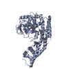
| Entry | Database: PDB / ID: 1zws | ||||||
|---|---|---|---|---|---|---|---|
| Title | Crystal structure of the catalytic domain of human DRP-1 kinase | ||||||
 Components Components | DAP-kinase related protein 1 | ||||||
 Keywords Keywords | TRANSFERASE / protein kinase / twinning | ||||||
| Function / homology |  Function and homology information Function and homology informationpositive regulation of eosinophil chemotaxis / autophagosome lumen / regulation of intrinsic apoptotic signaling pathway / neutrophil migration / Caspase activation via Dependence Receptors in the absence of ligand / anoikis / positive regulation of neutrophil chemotaxis / regulation of autophagy / protein autophosphorylation / cytoplasmic vesicle ...positive regulation of eosinophil chemotaxis / autophagosome lumen / regulation of intrinsic apoptotic signaling pathway / neutrophil migration / Caspase activation via Dependence Receptors in the absence of ligand / anoikis / positive regulation of neutrophil chemotaxis / regulation of autophagy / protein autophosphorylation / cytoplasmic vesicle / regulation of apoptotic process / protein phosphorylation / calmodulin binding / non-specific serine/threonine protein kinase / intracellular signal transduction / positive regulation of apoptotic process / protein serine kinase activity / protein serine/threonine kinase activity / apoptotic process / Golgi apparatus / ATP binding / identical protein binding / nucleus / cytoplasm Similarity search - Function | ||||||
| Biological species |  Homo sapiens (human) Homo sapiens (human) | ||||||
| Method |  X-RAY DIFFRACTION / SYNCHROTRON / MOLECULAR REPLACEMENT / Resolution: 2.9 Å X-RAY DIFFRACTION / SYNCHROTRON / MOLECULAR REPLACEMENT / Resolution: 2.9 Å | ||||||
 Authors Authors | Kursula, P. / Schunck, H. / Wilmanns, M. | ||||||
 Citation Citation | Journal: To be Published Title: Crystal structure of the catalytic domain of human DRP-1 kinase Authors: Kursula, P. / Schunck, H. / Wilmanns, M. | ||||||
| History |
|
- Structure visualization
| Structure viewer | Molecule: MolmilJmol/JSmol |
|---|
- Downloads & links
-Download
| PDBx/mmCIF format | 1zws.cif.gz | 407.7 KB | Display | PDBx/mmCIF format |
|---|---|---|---|---|
| PDB format | pdb1zws.ent.gz | 332.5 KB | Display | PDB format |
| PDBx/mmJSON format | 1zws.json.gz | Tree view | PDBx/mmJSON format | |
| Others |  Other downloads Other downloads |
-Validation report
| Arichive directory | https://data.pdbj.org/pub/pdb/validation_reports/zw/1zwsftp://data.pdbj.org/pub/pdb/validation_reports/zw/1zws | HTTPS FTP |
|---|
-Related structure data
| Related structure data |  1sxo S: Starting model for refinement |
|---|---|
| Similar structure data |


-Links
 PDBj
PDBj
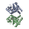
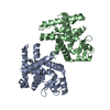
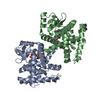
- Assembly
| Deposited unit | 
| ||||||||
|---|---|---|---|---|---|---|---|---|---|
| 1 | 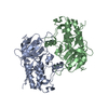
| ||||||||
| 2 | 
| ||||||||
| 3 | 
| ||||||||
| 4 | 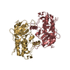
| ||||||||
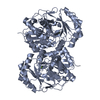
| Unit cell |
|
-Components
| #1: Protein | Mass: 33096.770 Da / Num. of mol.: 8 / Fragment: catalytic domain Source method: isolated from a genetically manipulated source Source: (gene. exp.) Homo sapiens (human) / Plasmid: pDEST-15 / Production host:  |
|---|
-Experimental details
-Experiment
| Experiment | Method: X-RAY DIFFRACTION / Number of used crystals: 1 |
|---|
- Sample preparation
| Crystal | Density Matthews: 2.7 Å3/Da / Density % sol: 54.7 % |
|---|---|
| Crystal grow | Temperature: 295 K / Method: vapor diffusion, hanging drop / pH: 7.5 Details: Tris, PEG, magnesium chloride, pH 7.5, VAPOR DIFFUSION, HANGING DROP, temperature 295K |
-Data collection
| Diffraction | Mean temperature: 100 K |
|---|---|
| Diffraction source | Source: SYNCHROTRON / Site: MAX II  / Beamline: I711 / Wavelength: 1.095 Å / Beamline: I711 / Wavelength: 1.095 Å |
| Detector | Type: MARRESEARCH / Detector: CCD / Date: Sep 27, 2004 |
| Radiation | Protocol: SINGLE WAVELENGTH / Monochromatic (M) / Laue (L): M / Scattering type: x-ray |
| Radiation wavelength | Wavelength: 1.095 Å / Relative weight: 1 |
| Reflection | Resolution: 2.9→20 Å / Num. all: 55013 / Num. obs: 55013 / % possible obs: 90.5 % / Observed criterion σ(F): -3 / Observed criterion σ(I): -3 / Redundancy: 1.9 % / Biso Wilson estimate: 48 Å2 / Rsym value: 0.115 / Net I/σ(I): 6.1 |
| Reflection shell | Resolution: 2.9→3 Å / Redundancy: 1.6 % / Mean I/σ(I) obs: 1.5 / Num. unique all: 4903 / Rsym value: 0.503 / % possible all: 84.2 |
- Processing
| Software |
| |||||||||||||||||||||||||
|---|---|---|---|---|---|---|---|---|---|---|---|---|---|---|---|---|---|---|---|---|---|---|---|---|---|---|
| Refinement | Method to determine structure: MOLECULAR REPLACEMENT Starting model: 1sxo 1sxo Resolution: 2.9→20 Å / σ(F): -3 / Stereochemistry target values: Engh & Huber Details: The crystal form is perfectly twinned with the operator -h,-k,h+l, twinning fraction = 0.5
| |||||||||||||||||||||||||
| Refinement step | Cycle: LAST / Resolution: 2.9→20 Å
| |||||||||||||||||||||||||
| Refine LS restraints |
|
