 Movie
Movie Controller


+ Open data

- Basic information
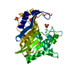
| Entry | Database: PDB / ID: 1znc | ||||||
|---|---|---|---|---|---|---|---|
| Title | HUMAN CARBONIC ANHYDRASE IV | ||||||
 Components Components | CARBONIC ANHYDRASE IV | ||||||
 Keywords Keywords | LYASE / GPI-ANCHOR / MEMBRANE / ZINC | ||||||
| Function / homology |  Function and homology information Function and homology informationbicarbonate transport / transport vesicle membrane / endoplasmic reticulum-Golgi intermediate compartment / rough endoplasmic reticulum / Reversible hydration of carbon dioxide / secretory granule membrane / carbonic anhydrase / carbonate dehydratase activity / brush border membrane / trans-Golgi network ...bicarbonate transport / transport vesicle membrane / endoplasmic reticulum-Golgi intermediate compartment / rough endoplasmic reticulum / Reversible hydration of carbon dioxide / secretory granule membrane / carbonic anhydrase / carbonate dehydratase activity / brush border membrane / trans-Golgi network / Erythrocytes take up oxygen and release carbon dioxide / Erythrocytes take up carbon dioxide and release oxygen / apical plasma membrane / external side of plasma membrane / perinuclear region of cytoplasm / cell surface / Golgi apparatus / extracellular exosome / zinc ion binding / membrane / plasma membrane Similarity search - Function | ||||||
| Biological species |  Homo sapiens (human) Homo sapiens (human) | ||||||
| Method |  X-RAY DIFFRACTION / MOLECULAR REPLACEMENT / Resolution: 2.8 Å X-RAY DIFFRACTION / MOLECULAR REPLACEMENT / Resolution: 2.8 Å | ||||||
 Authors Authors | Stams, T. / Christianson, D.W. | ||||||
 Citation Citation | Journal: Proc.Natl.Acad.Sci.USA / Year: 1996 Title: Crystal structure of the secretory form of membrane-associated human carbonic anhydrase IV at 2.8-A resolution. Authors: Stams, T. / Nair, S.K. / Okuyama, T. / Waheed, A. / Sly, W.S. / Christianson, D.W. #1: Journal: Arch.Biochem.Biophys. / Year: 1996Title: Carbonic Anhydrase Iv: Purification of a Secretory Form of the Recombinant Human Enzyme and Identification of the Positions and Importance of its Disulfide Bonds Authors: Waheed, A. / Okuyama, T. / Heyduk, T. / Sly, W.S. | ||||||
| History |
|
- Structure visualization
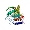
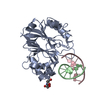
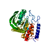
| Structure viewer | Molecule: MolmilJmol/JSmol |
|---|
- Downloads & links
-Download
| PDBx/mmCIF format | 1znc.cif.gz | 131.7 KB | Display | PDBx/mmCIF format |
|---|---|---|---|---|
| PDB format | pdb1znc.ent.gz | 102.9 KB | Display | PDB format |
| PDBx/mmJSON format | 1znc.json.gz | Tree view | PDBx/mmJSON format | |
| Others |  Other downloads Other downloads |
-Validation report
| Arichive directory | https://data.pdbj.org/pub/pdb/validation_reports/zn/1zncftp://data.pdbj.org/pub/pdb/validation_reports/zn/1znc | HTTPS FTP |
|---|
-Related structure data
| Similar structure data |
|---|


-Links
 PDBj
PDBj- Assembly
| Deposited unit | 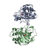
| ||||||||
|---|---|---|---|---|---|---|---|---|---|
| 1 | 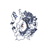
| ||||||||
| 2 | 
| ||||||||
| Unit cell |
|
-Components
| #1: Protein | Mass: 30364.473 Da / Num. of mol.: 2 Source method: isolated from a genetically manipulated source Source: (gene. exp.) Homo sapiens (human) / Gene: HUMAN CAIV / Plasmid: PCAGGS / Cell line (production host): CHO-CELLS / Production host:   Cricetulus griseus (Chinese hamster) / References: UniProt: P22748, carbonic anhydrase Cricetulus griseus (Chinese hamster) / References: UniProt: P22748, carbonic anhydrase#2: Chemical |   Mass: 96.063 Da / Num. of mol.: 2 / Source method: obtained synthetically / Formula: SO4 Mass: 96.063 Da / Num. of mol.: 2 / Source method: obtained synthetically / Formula: SO4#3: Chemical |   Mass: 65.409 Da / Num. of mol.: 2 / Source method: obtained synthetically / Formula: Zn Mass: 65.409 Da / Num. of mol.: 2 / Source method: obtained synthetically / Formula: Zn#4: Water | ChemComp-HOH / |  Mass: 18.015 Da / Num. of mol.: 32 / Source method: isolated from a natural source / Formula: H2O Mass: 18.015 Da / Num. of mol.: 32 / Source method: isolated from a natural source / Formula: H2OHas protein modification | Y | Sequence details | THE RESIDUE NUMBERING IS BASED ON THE RESIDUE NUMBERING OF CARBONIC ANHYDRASE II. INSERTIONS ARE ...THE RESIDUE NUMBERING IS BASED ON THE RESIDUE NUMBERING OF CARBONIC ANHYDRASE II. INSERTIONS | |
|---|
-Experimental details
-Experiment
| Experiment | Method: X-RAY DIFFRACTION / Number of used crystals: 2 |
|---|
- Sample preparation
| Crystal | Density Matthews: 2.4 Å3/Da / Density % sol: 48.9 % | ||||||||||||||||||||||||||||||||||||||||||
|---|---|---|---|---|---|---|---|---|---|---|---|---|---|---|---|---|---|---|---|---|---|---|---|---|---|---|---|---|---|---|---|---|---|---|---|---|---|---|---|---|---|---|---|
| Crystal grow | pH: 5.1 / Details: 25% PEG 3,350; 100MM NA ACETATE PH 5.1 | ||||||||||||||||||||||||||||||||||||||||||
| Crystal grow | *PLUS Method: vapor diffusion, hanging drop | ||||||||||||||||||||||||||||||||||||||||||
| Components of the solutions | *PLUS
|
-Data collection
| Diffraction | Mean temperature: 298 K |
|---|---|
| Diffraction source | Source: ROTATING ANODE / Type: RIGAKU RUH2R / Wavelength: 1.5418 |
| Detector | Type: RIGAKU / Detector: IMAGE PLATE / Details: MIRRORS |
| Radiation | Monochromatic (M) / Laue (L): M / Scattering type: x-ray |
| Radiation wavelength | Wavelength: 1.5418 Å / Relative weight: 1 |
| Reflection | Resolution: 2.8→20 Å / Num. obs: 13626 / % possible obs: 95 % / Redundancy: 3.4 % / Rmerge(I) obs: 0.08 |
| Reflection shell | Resolution: 2.8→2.87 Å / Redundancy: 3.3 % / Rmerge(I) obs: 0.253 / % possible all: 91.8 |
| Reflection | *PLUS Num. measured all: 46250 |
| Reflection shell | *PLUS % possible obs: 91.8 % |
- Processing
| Software |
| ||||||||||||||||||||||||||||||||||||||||||||||||||||||||||||
|---|---|---|---|---|---|---|---|---|---|---|---|---|---|---|---|---|---|---|---|---|---|---|---|---|---|---|---|---|---|---|---|---|---|---|---|---|---|---|---|---|---|---|---|---|---|---|---|---|---|---|---|---|---|---|---|---|---|---|---|---|---|
| Refinement | Method to determine structure: MOLECULAR REPLACEMENT Starting model: POLYALANINE MODEL OF CAII MISSING N-TERMINAL 30 Resolution: 2.8→20 Å / Cross valid method: FREE-R / σ(F): 2
| ||||||||||||||||||||||||||||||||||||||||||||||||||||||||||||
| Displacement parameters | Biso mean: 28.3 Å2 | ||||||||||||||||||||||||||||||||||||||||||||||||||||||||||||
| Refinement step | Cycle: LAST / Resolution: 2.8→20 Å
| ||||||||||||||||||||||||||||||||||||||||||||||||||||||||||||
| Refine LS restraints |
| ||||||||||||||||||||||||||||||||||||||||||||||||||||||||||||
| Refine LS restraints NCS | Rms dev position: 0.2 Å | ||||||||||||||||||||||||||||||||||||||||||||||||||||||||||||
| LS refinement shell | Resolution: 2.8→2.93 Å
| ||||||||||||||||||||||||||||||||||||||||||||||||||||||||||||
| Software | *PLUS Name: X-PLOR / Version: 3.1 / Classification: refinement | ||||||||||||||||||||||||||||||||||||||||||||||||||||||||||||
| Refinement | *PLUS | ||||||||||||||||||||||||||||||||||||||||||||||||||||||||||||
| Solvent computation | *PLUS | ||||||||||||||||||||||||||||||||||||||||||||||||||||||||||||
| Displacement parameters | *PLUS | ||||||||||||||||||||||||||||||||||||||||||||||||||||||||||||
| Refine LS restraints | *PLUS
| ||||||||||||||||||||||||||||||||||||||||||||||||||||||||||||
| LS refinement shell | *PLUS Rfactor obs: 0.28 |
