 Movie
Movie Controller

[English] 日本語
 Yorodumi
- PDB-1zmt: Structure of haloalcohol dehalogenase HheC of Agrobacterium radio... -
+ Open data

- Basic information
| Entry | Database: PDB / ID: 1zmt | ||||||
|---|---|---|---|---|---|---|---|
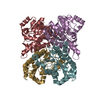
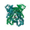
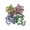
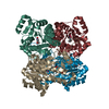
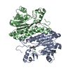
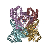
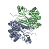
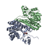
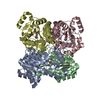
| Title | Structure of haloalcohol dehalogenase HheC of Agrobacterium radiobacter AD1 in complex with (R)-para-nitro styrene oxide, with a water molecule in the halide-binding site | ||||||
 Components Components | Haloalcohol dehalogenase HheC | ||||||
 Keywords Keywords | LYASE / haloalcohol dehalogenase / halohydrin dehalogenase / epoxide catalysis / enantioselectivity | ||||||
| Function / homology |  Function and homology information Function and homology information: / Enoyl-(Acyl carrier protein) reductase / Short-chain dehydrogenase/reductase SDR / NAD(P)-binding Rossmann-like Domain / NAD(P)-binding domain superfamily / Rossmann fold / 3-Layer(aba) Sandwich / Alpha Beta Similarity search - Domain/homology | ||||||
| Biological species |  Agrobacterium tumefaciens (bacteria) Agrobacterium tumefaciens (bacteria) | ||||||
| Method |  X-RAY DIFFRACTION / SYNCHROTRON / MOLECULAR REPLACEMENT / Resolution: 1.7 Å X-RAY DIFFRACTION / SYNCHROTRON / MOLECULAR REPLACEMENT / Resolution: 1.7 Å | ||||||
 Authors Authors | de Jong, R.M. / Tiesinga, J.J.W. / Villa, A. / Tang, L. / Janssen, D.B. / Dijkstra, B.W. | ||||||
 Citation Citation | Journal: J.Am.Chem.Soc. / Year: 2005 Title: Structural Basis for the Enantioselectivity of an Epoxide Ring Opening Reaction Catalyzed by Halo Alcohol Dehalogenase HheC. Authors: de Jong, R.M. / Tiesinga, J.J.W. / Villa, A. / Tang, L. / Janssen, D.B. / Dijkstra, B.W. | ||||||
| History |
|
- Structure visualization
| Structure viewer | Molecule: MolmilJmol/JSmol |
|---|
- Downloads & links
-Download
| PDBx/mmCIF format | 1zmt.cif.gz | 211.8 KB | Display | PDBx/mmCIF format |
|---|---|---|---|---|
| PDB format | pdb1zmt.ent.gz | 169.2 KB | Display | PDB format |
| PDBx/mmJSON format | 1zmt.json.gz | Tree view | PDBx/mmJSON format | |
| Others |  Other downloads Other downloads |
-Validation report
| Arichive directory | https://data.pdbj.org/pub/pdb/validation_reports/zm/1zmtftp://data.pdbj.org/pub/pdb/validation_reports/zm/1zmt | HTTPS FTP |
|---|
-Related structure data


-Links
 PDBj
PDBj

- Assembly
| Deposited unit | 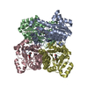
| ||||||||
|---|---|---|---|---|---|---|---|---|---|
| 1 |
| ||||||||
| Unit cell |
| ||||||||
| Details | The asymmetric unit contains two dimers that form two biologically relevant tetramers |
-Components
| #1: Protein | Mass: 27979.666 Da / Num. of mol.: 4 Source method: isolated from a genetically manipulated source Source: (gene. exp.) Agrobacterium tumefaciens (bacteria) / Strain: AD1 / Plasmid: pBADHheC / Species (production host): Escherichia coli / Production host: #2: Chemical | ChemComp-RNO / (   Mass: 165.146 Da / Num. of mol.: 4 / Source method: obtained synthetically / Formula: C8H7NO3 Mass: 165.146 Da / Num. of mol.: 4 / Source method: obtained synthetically / Formula: C8H7NO3#3: Water | ChemComp-HOH / |  Mass: 18.015 Da / Num. of mol.: 716 / Source method: isolated from a natural source / Formula: H2O Mass: 18.015 Da / Num. of mol.: 716 / Source method: isolated from a natural source / Formula: H2O |
|---|
-Experimental details
-Experiment
| Experiment | Method: X-RAY DIFFRACTION / Number of used crystals: 1 |
|---|
- Sample preparation
| Crystal | Density Matthews: 2.37 Å3/Da / Density % sol: 47.8 % |
|---|---|
| Crystal grow | Temperature: 298 K / Method: vapor diffusion, hanging drop / pH: 6.9 Details: 100 mM bis-Tris buffer, 50% Ammonium sulfate, pH 6.9, VAPOR DIFFUSION, HANGING DROP, temperature 298K |
-Data collection
| Diffraction | Mean temperature: 100 K |
|---|---|
| Diffraction source | Source: SYNCHROTRON / Site: ESRF  / Beamline: ID14-2 / Wavelength: 0.93 Å / Beamline: ID14-2 / Wavelength: 0.93 Å |
| Detector | Type: ADSC QUANTUM 4 / Detector: CCD / Date: Apr 9, 2003 |
| Radiation | Monochromator: Si / Protocol: SINGLE WAVELENGTH / Monochromatic (M) / Laue (L): M / Scattering type: x-ray |
| Radiation wavelength | Wavelength: 0.93 Å / Relative weight: 1 |
| Reflection | Resolution: 1.7→30 Å / Num. all: 112897 / Num. obs: 110598 / % possible obs: 99.4 % / Observed criterion σ(F): 2 / Observed criterion σ(I): 2 / Biso Wilson estimate: 18.9 Å2 |
| Reflection shell | Resolution: 1.7→1.81 Å / % possible all: 95.6 |
- Processing
| Software |
| ||||||||||||||||||||||||||||||||||||
|---|---|---|---|---|---|---|---|---|---|---|---|---|---|---|---|---|---|---|---|---|---|---|---|---|---|---|---|---|---|---|---|---|---|---|---|---|---|
| Refinement | Method to determine structure: MOLECULAR REPLACEMENT / Resolution: 1.7→27.2 Å / Rfactor Rfree error: 0.002 / Data cutoff high absF: 2431700.55 / Data cutoff high rms absF: 2431700.55 / Data cutoff low absF: 0 / Isotropic thermal model: RESTRAINED / Cross valid method: THROUGHOUT / σ(F): 0 / Stereochemistry target values: Engh & Huber
| ||||||||||||||||||||||||||||||||||||
| Solvent computation | Solvent model: FLAT MODEL / Bsol: 59.6245 Å2 / ksol: 0.425254 e/Å3 | ||||||||||||||||||||||||||||||||||||
| Displacement parameters | Biso mean: 18 Å2
| ||||||||||||||||||||||||||||||||||||
| Refine analyze |
| ||||||||||||||||||||||||||||||||||||
| Refinement step | Cycle: LAST / Resolution: 1.7→27.2 Å
| ||||||||||||||||||||||||||||||||||||
| Refine LS restraints |
| ||||||||||||||||||||||||||||||||||||
| LS refinement shell | Resolution: 1.7→1.81 Å / Rfactor Rfree error: 0.008 / Total num. of bins used: 6
| ||||||||||||||||||||||||||||||||||||
| Xplor file |
|
