 Movie
Movie Controller

[English] 日本語
 Yorodumi
- PDB-1zk2: Orthorhombic crystal structure of the apo-form of R-specific alco... -
+ Open data

- Basic information
| Entry | Database: PDB / ID: 1zk2 | ||||||
|---|---|---|---|---|---|---|---|
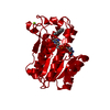
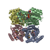
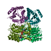
| Title | Orthorhombic crystal structure of the apo-form of R-specific alcohol dehydrogenase (mutant G37D) from Lactobacillus brevis | ||||||
 Components Components | R-specific alcohol dehydrogenase | ||||||
 Keywords Keywords | OXIDOREDUCTASE / short chain reductases/dehydrogenases / magnesium dependence / R-specific alcohol dehydrogenase | ||||||
| Function / homology |  Function and homology information Function and homology informationbile acid metabolic process / oxidoreductase activity / nucleotide binding / metal ion binding Similarity search - Function | ||||||
| Biological species |  Lactobacillus brevis (bacteria) Lactobacillus brevis (bacteria) | ||||||
| Method |  X-RAY DIFFRACTION / MOLECULAR REPLACEMENT / Resolution: 1.55 Å X-RAY DIFFRACTION / MOLECULAR REPLACEMENT / Resolution: 1.55 Å | ||||||
 Authors Authors | Schlieben, N.H. / Niefind, K. / Muller, J. / Riebel, B. / Hummel, W. / Schomburg, D. | ||||||
 Citation Citation | Journal: J.Mol.Biol. / Year: 2005 Title: Atomic resolution structures of R-specific alcohol dehydrogenase from Lactobacillus brevis provide the structural bases of its substrate and cosubstrate specificity Authors: Schlieben, N.H. / Niefind, K. / Muller, J. / Riebel, B. / Hummel, W. / Schomburg, D. #1: Journal: J.Mol.Biol. / Year: 2003Title: The crystal structure of R-specific alcohol dehydrogenase from Lactobacillus brevis suggests the structural basis of its metal dependency Authors: Niefind, K. / Muller, J. / Riebel, B. / Hummel, W. / Schomburg, D. #2: Journal: Acta Crystallogr.,Sect.D / Year: 2000 Title: Crystallization and preliminary characterization of crystals of R-alcohol dehydrogenase from Lactobacills brevis Authors: Niefind, K. / Riebel, B. / Muller, J. / Hummel, W. / Schomburg, D. | ||||||
| History |
|
- Structure visualization
| Structure viewer | Molecule: MolmilJmol/JSmol |
|---|
- Downloads & links
-Download
| PDBx/mmCIF format | 1zk2.cif.gz | 74.8 KB | Display | PDBx/mmCIF format |
|---|---|---|---|---|
| PDB format | pdb1zk2.ent.gz | 54.7 KB | Display | PDB format |
| PDBx/mmJSON format | 1zk2.json.gz | Tree view | PDBx/mmJSON format | |
| Others |  Other downloads Other downloads |
-Validation report
| Arichive directory | https://data.pdbj.org/pub/pdb/validation_reports/zk/1zk2ftp://data.pdbj.org/pub/pdb/validation_reports/zk/1zk2 | HTTPS FTP |
|---|
-Related structure data
| Related structure data |  1zjyC  1zjzC  1zk0C  1zk1C 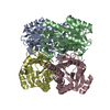 1zk3C  1zk4C C: citing same article ( |
|---|---|
| Similar structure data |


-Links
 PDBj
PDBj


- Assembly
| Deposited unit | 
| |||||||||||||||
|---|---|---|---|---|---|---|---|---|---|---|---|---|---|---|---|---|
| 1 | 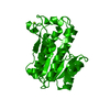
| |||||||||||||||
| Unit cell |
| |||||||||||||||
| Components on special symmetry positions |
| |||||||||||||||
| Details | The biological assembly is a tetramer generated form the monomer in the asymmetric unit by the following operations: x, y, z; -x, -y, z; -x, y, -z; x, -y, -z |
-Components
| #1: Protein | Mass: 26714.098 Da / Num. of mol.: 1 / Mutation: G37D Source method: isolated from a genetically manipulated source Source: (gene. exp.) Lactobacillus brevis (bacteria) / Plasmid: pkk177-3H / Production host: |
|---|---|
| #2: Chemical | ChemComp-MG /   Mass: 24.305 Da / Num. of mol.: 1 / Source method: obtained synthetically / Formula: Mg Mass: 24.305 Da / Num. of mol.: 1 / Source method: obtained synthetically / Formula: Mg |
| #3: Water | ChemComp-HOH /  Mass: 18.015 Da / Num. of mol.: 429 / Source method: isolated from a natural source / Formula: H2O Mass: 18.015 Da / Num. of mol.: 429 / Source method: isolated from a natural source / Formula: H2O |
-Experimental details
-Experiment
| Experiment | Method: X-RAY DIFFRACTION / Number of used crystals: 1 |
|---|
- Sample preparation
| Crystal | Density Matthews: 2.4 Å3/Da / Density % sol: 47.5 % |
|---|
-Data collection
| Diffraction source | Source: ROTATING ANODE / Type: ENRAF-NONIUS FR571 / Wavelength: 1.5418 Å |
|---|---|
| Radiation | Protocol: SINGLE WAVELENGTH / Monochromatic (M) / Laue (L): M / Scattering type: x-ray |
| Radiation wavelength | Wavelength: 1.5418 Å / Relative weight: 1 |
| Reflection | Resolution: 1.55→65.3 Å / Num. all: 37100 / Num. obs: 36602 |
- Processing
| Software |
| ||||||||||||||||||||||||||||||||||||||||||||||||||||||||||||||||||||||||||||||||||||||||||||||||||||||||||||||||||||||||||||||||||||||||||||||||||||||||||||||||||||||||||
|---|---|---|---|---|---|---|---|---|---|---|---|---|---|---|---|---|---|---|---|---|---|---|---|---|---|---|---|---|---|---|---|---|---|---|---|---|---|---|---|---|---|---|---|---|---|---|---|---|---|---|---|---|---|---|---|---|---|---|---|---|---|---|---|---|---|---|---|---|---|---|---|---|---|---|---|---|---|---|---|---|---|---|---|---|---|---|---|---|---|---|---|---|---|---|---|---|---|---|---|---|---|---|---|---|---|---|---|---|---|---|---|---|---|---|---|---|---|---|---|---|---|---|---|---|---|---|---|---|---|---|---|---|---|---|---|---|---|---|---|---|---|---|---|---|---|---|---|---|---|---|---|---|---|---|---|---|---|---|---|---|---|---|---|---|---|---|---|---|---|---|---|
| Refinement | Method to determine structure: MOLECULAR REPLACEMENT / Resolution: 1.55→65.3 Å / Cor.coef. Fo:Fc: 0.969 / Cor.coef. Fo:Fc free: 0.958 / SU B: 1.185 / SU ML: 0.043 / Cross valid method: THROUGHOUT / σ(F): 0 / ESU R: 0.073 / ESU R Free: 0.072 / Stereochemistry target values: MAXIMUM LIKELIHOOD / Details: HYDROGENS HAVE BEEN ADDED IN THE RIDING POSITIONS
| ||||||||||||||||||||||||||||||||||||||||||||||||||||||||||||||||||||||||||||||||||||||||||||||||||||||||||||||||||||||||||||||||||||||||||||||||||||||||||||||||||||||||||
| Solvent computation | Ion probe radii: 0.8 Å / Shrinkage radii: 0.8 Å / VDW probe radii: 1.4 Å / Solvent model: BABINET MODEL WITH MASK | ||||||||||||||||||||||||||||||||||||||||||||||||||||||||||||||||||||||||||||||||||||||||||||||||||||||||||||||||||||||||||||||||||||||||||||||||||||||||||||||||||||||||||
| Displacement parameters | Biso mean: 12.886 Å2
| ||||||||||||||||||||||||||||||||||||||||||||||||||||||||||||||||||||||||||||||||||||||||||||||||||||||||||||||||||||||||||||||||||||||||||||||||||||||||||||||||||||||||||
| Refinement step | Cycle: LAST / Resolution: 1.55→65.3 Å
| ||||||||||||||||||||||||||||||||||||||||||||||||||||||||||||||||||||||||||||||||||||||||||||||||||||||||||||||||||||||||||||||||||||||||||||||||||||||||||||||||||||||||||
| Refine LS restraints |
| ||||||||||||||||||||||||||||||||||||||||||||||||||||||||||||||||||||||||||||||||||||||||||||||||||||||||||||||||||||||||||||||||||||||||||||||||||||||||||||||||||||||||||
| LS refinement shell | Resolution: 1.55→1.59 Å / Total num. of bins used: 20 /
|
