 Movie
Movie Controller


+ Open data

- Basic information
| Entry | Database: PDB / ID: 1zbb | ||||||
|---|---|---|---|---|---|---|---|
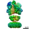
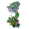
| Title | Structure of the 4_601_167 Tetranucleosome | ||||||
 Components Components |
| ||||||
 Keywords Keywords | STRUCTURAL PROTEIN/DNA / TETRANUCLEOSOME / NUCLEOSOME / CHROMATIN / CHROMATIN FIBER / HISTONE / PROTEIN-DNA INTERACTION / NUCLEOPROTEIN / SUPERCOILED DNA / NUCLEOSOME CORE / PROTEIN-DNA COMPLEX / STRUCTURAL PROTEIN-DNA COMPLEX | ||||||
| Function / homology |  Function and homology information Function and homology informationstructural constituent of chromatin / nucleosome / nucleosome assembly / heterochromatin formation / protein heterodimerization activity / DNA binding / nucleoplasm / nucleus Similarity search - Function | ||||||
| Biological species | |||||||
| Method |  X-RAY DIFFRACTION / SYNCHROTRON / MOLECULAR REPLACEMENT / Resolution: 9 Å X-RAY DIFFRACTION / SYNCHROTRON / MOLECULAR REPLACEMENT / Resolution: 9 Å | ||||||
 Authors Authors | Schalch, T. / Duda, S. / Sargent, D.F. / Richmond, T.J. | ||||||
 Citation Citation | Journal: Nature / Year: 2005 Title: X-ray structure of a tetranucleosome and its implications for the chromatin fibre. Authors: Schalch, T. / Duda, S. / Sargent, D.F. / Richmond, T.J. | ||||||
| History |
|
- Structure visualization
| Structure viewer | Molecule: MolmilJmol/JSmol |
|---|
- Downloads & links
-Download
| PDBx/mmCIF format | 1zbb.cif.gz | 605.9 KB | Display | PDBx/mmCIF format |
|---|---|---|---|---|
| PDB format | pdb1zbb.ent.gz | 454 KB | Display | PDB format |
| PDBx/mmJSON format | 1zbb.json.gz | Tree view | PDBx/mmJSON format | |
| Others |  Other downloads Other downloads |
-Validation report
| Arichive directory | https://data.pdbj.org/pub/pdb/validation_reports/zb/1zbbftp://data.pdbj.org/pub/pdb/validation_reports/zb/1zbb | HTTPS FTP |
|---|
-Related structure data
| Related structure data | 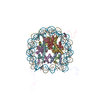 1kx5S S: Starting model for refinement |
|---|---|
| Similar structure data |


-Links
 PDBj
PDBj


- Assembly
| Deposited unit | 
| ||||||||
|---|---|---|---|---|---|---|---|---|---|
| 1 | 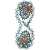
| ||||||||
| Unit cell |
| ||||||||
| Details | The second part of the biological assembly is generated by the two fold axis: -X+1,Y,-Z+1 |
-Components
-DNA chain , 2 types, 2 molecules IJ
| #1: DNA chain | Mass: 107153.336 Da / Num. of mol.: 1 / Source method: obtained synthetically Details: Tandem repeat of sequence isolated by Lowary PT, Widom J., J Mol Biol. 1998 Feb 13;276(1):19-42 SEE REMARK 999 for more details |
|---|---|
| #2: DNA chain | Mass: 107144.312 Da / Num. of mol.: 1 / Source method: obtained synthetically Details: Tandem repeat of sequence isolated by Lowary PT, Widom J., J Mol Biol. 1998 Feb 13;276(1):19-42 SEE REMARK 999 for more details |
-Protein , 4 types, 16 molecules AEaeBFbfCGcgDHdh
| #3: Protein | Mass: 15303.930 Da / Num. of mol.: 4 Source method: isolated from a genetically manipulated source Source: (gene. exp.)  #4: Protein | Mass: 11263.231 Da / Num. of mol.: 4 Source method: isolated from a genetically manipulated source Source: (gene. exp.) #5: Protein | Mass: 13978.241 Da / Num. of mol.: 4 Source method: isolated from a genetically manipulated source Source: (gene. exp.) #6: Protein | Mass: 13848.097 Da / Num. of mol.: 4 Source method: isolated from a genetically manipulated source Source: (gene. exp.) |
|---|
-Details
| Has protein modification | Y |
|---|---|
| Sequence details | THE 4_601_167 TETRANUCLEOSOME IS MODELLED BASED ON THE NUCLEOSOME CORE PARTICLE STRUCTURE 1KX5. DUE ...THE 4_601_167 TETRANUCLE |
-Experimental details
-Experiment
| Experiment | Method: X-RAY DIFFRACTION / Number of used crystals: 12 |
|---|
- Sample preparation
| Crystal | Density Matthews: 2.9 Å3/Da / Density % sol: 57.2 % | ||||||||||||||||||||||||||||||||||||
|---|---|---|---|---|---|---|---|---|---|---|---|---|---|---|---|---|---|---|---|---|---|---|---|---|---|---|---|---|---|---|---|---|---|---|---|---|---|
| Crystal grow | Temperature: 294 K / Method: vapor diffusion, sitting drop / pH: 6.75 Details: magnesium chloride, potassium chloride, potassium cacodylate, trisCl, pH 6.75, VAPOR DIFFUSION, SITTING DROP, temperature 294K | ||||||||||||||||||||||||||||||||||||
| Components of the solutions |
|
-Data collection
| Diffraction |
| ||||||||||||||||||||||||
|---|---|---|---|---|---|---|---|---|---|---|---|---|---|---|---|---|---|---|---|---|---|---|---|---|---|
| Diffraction source |
| ||||||||||||||||||||||||
| Detector |
| ||||||||||||||||||||||||
| Radiation |
| ||||||||||||||||||||||||
| Radiation wavelength |
| ||||||||||||||||||||||||
| Reflection | Resolution: 9→50 Å / Num. all: 1994 / Num. obs: 1994 / % possible obs: 97 % / Observed criterion σ(F): 0 / Observed criterion σ(I): 0 / Redundancy: 5.7 % / Rsym value: 0.072 / Net I/σ(I): 17.28 | ||||||||||||||||||||||||
| Reflection shell | Resolution: 9→10 Å / Redundancy: 4.2 % / Mean I/σ(I) obs: 12.55 / Num. unique all: 542 / Rsym value: 0.087 / % possible all: 97.4 |

- Processing
| Software |
| ||||||||||||||||
|---|---|---|---|---|---|---|---|---|---|---|---|---|---|---|---|---|---|
| Refinement | Method to determine structure: MOLECULAR REPLACEMENT Starting model: 1KX5(NUCLEOSOME CORE PARTICLE) Resolution: 9→50 Å / σ(F): 0 Details: THE STRUCTURE WAS REFINED BY RIGID BODY REFINEMENT OF NUCLEOSOME POSITIONS ONLY. NO CROSSVALIDATION WAS EMPLOYED DUE TO HIGH FLUCTUATIONS OF RFREE CAUSED BY THE SMALL NUMBER OF REFLECTIONS ...Details: THE STRUCTURE WAS REFINED BY RIGID BODY REFINEMENT OF NUCLEOSOME POSITIONS ONLY. NO CROSSVALIDATION WAS EMPLOYED DUE TO HIGH FLUCTUATIONS OF RFREE CAUSED BY THE SMALL NUMBER OF REFLECTIONS IN THE TEST SET. Since the structure was solved at very low resolution, remark 500 records have been suppressed at the request of depositors.
| ||||||||||||||||
| Refinement step | Cycle: LAST / Resolution: 9→50 Å
| ||||||||||||||||
| LS refinement shell | Resolution: 9→9.72 Å
|
