 Movie
Movie Controller


+ Open data

- Basic information
| Entry | Database: PDB / ID: 1z7e | ||||||
|---|---|---|---|---|---|---|---|
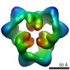
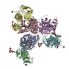
| Title | Crystal structure of full length ArnA | ||||||
 Components Components | protein ArnA | ||||||
 Keywords Keywords | HYDROLASE / Rossmann fold / OB-like fold | ||||||
| Function / homology |  Function and homology information Function and homology informationUDP-4-deoxy-4-formamido-beta-L-arabinopyranose biosynthetic process / UDP-glucuronate dehydrogenase activity / UDP-4-amino-4-deoxy-L-arabinose formyltransferase activity / UDP-glucuronic acid dehydrogenase (UDP-4-keto-hexauronic acid decarboxylating) / UDP-4-amino-4-deoxy-L-arabinose formyltransferase / UDP-D-xylose biosynthetic process / UDP-glucuronate decarboxylase activity / lipopolysaccharide biosynthetic process / lipid A biosynthetic process / NAD+ binding ...UDP-4-deoxy-4-formamido-beta-L-arabinopyranose biosynthetic process / UDP-glucuronate dehydrogenase activity / UDP-4-amino-4-deoxy-L-arabinose formyltransferase activity / UDP-glucuronic acid dehydrogenase (UDP-4-keto-hexauronic acid decarboxylating) / UDP-4-amino-4-deoxy-L-arabinose formyltransferase / UDP-D-xylose biosynthetic process / UDP-glucuronate decarboxylase activity / lipopolysaccharide biosynthetic process / lipid A biosynthetic process / NAD+ binding / response to antibiotic / protein-containing complex / membrane / identical protein binding Similarity search - Function | ||||||
| Biological species |  | ||||||
| Method |  X-RAY DIFFRACTION / SYNCHROTRON / MOLECULAR REPLACEMENT / Resolution: 3 Å X-RAY DIFFRACTION / SYNCHROTRON / MOLECULAR REPLACEMENT / Resolution: 3 Å | ||||||
 Authors Authors | Gatzeva-Topalova, P.Z. / May, A.P. / Sousa, M.C. | ||||||
 Citation Citation | Journal: Structure / Year: 2005 Title: Structure and Mechanism of ArnA: Conformational Change Implies Ordered Dehydrogenase Mechanism in Key Enzyme for Polymyxin Resistance Authors: Gatzeva-Topalova, P.Z. / May, A.P. / Sousa, M.C. | ||||||
| History |
|
- Structure visualization
| Structure viewer | Molecule: MolmilJmol/JSmol |
|---|
- Downloads & links
-Download
| PDBx/mmCIF format | 1z7e.cif.gz | 738.3 KB | Display | PDBx/mmCIF format |
|---|---|---|---|---|
| PDB format | pdb1z7e.ent.gz | 607.9 KB | Display | PDB format |
| PDBx/mmJSON format | 1z7e.json.gz | Tree view | PDBx/mmJSON format | |
| Others |  Other downloads Other downloads |
-Validation report
| Arichive directory | https://data.pdbj.org/pub/pdb/validation_reports/z7/1z7eftp://data.pdbj.org/pub/pdb/validation_reports/z7/1z7e | HTTPS FTP |
|---|
-Related structure data
| Related structure data |  1z73C  1z74C  1z75C  1z7bC  1u9jS 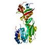 1yrwS C: citing same article ( S: Starting model for refinement |
|---|---|
| Similar structure data |
-Links
 PDBj
PDBj- Assembly
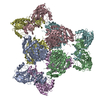
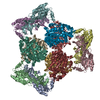
| Deposited unit | 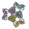
| ||||||||||
|---|---|---|---|---|---|---|---|---|---|---|---|
| 1 |
| ||||||||||
| Unit cell |
|
-Components
| #1: Protein | Mass: 74385.773 Da / Num. of mol.: 6 Source method: isolated from a genetically manipulated source Details: full length / Source: (gene. exp.) #2: Chemical | ChemComp-ATP /   Mass: 507.181 Da / Num. of mol.: 6 / Source method: obtained synthetically / Formula: C10H16N5O13P3 / Comment: ATP, energy-carrying molecule*YM Mass: 507.181 Da / Num. of mol.: 6 / Source method: obtained synthetically / Formula: C10H16N5O13P3 / Comment: ATP, energy-carrying molecule*YM#3: Chemical | ChemComp-UGA /   Mass: 580.285 Da / Num. of mol.: 6 / Source method: obtained synthetically / Formula: C15H22N2O18P2 Mass: 580.285 Da / Num. of mol.: 6 / Source method: obtained synthetically / Formula: C15H22N2O18P2 |
|---|
-Experimental details
-Experiment
| Experiment | Method: X-RAY DIFFRACTION / Number of used crystals: 1 |
|---|
- Sample preparation
| Crystal | Density Matthews: 3.7 Å3/Da / Density % sol: 67 % |
|---|---|
| Crystal grow | Temperature: 289 K / Method: vapor diffusion, hanging drop / pH: 7 Details: 0.1M MES pH 7.0, 10% Ethylene glycol, 9% PEG 8000, 14 mM 2mercaptoethanol and 10 mM ATP; protein was pre-incubated on ice with 3mM UDP-GlcA and 3 mM MgSO4 , VAPOR DIFFUSION, HANGING DROP, temperature 289K |
-Data collection
| Diffraction | Mean temperature: 100 K |
|---|---|
| Diffraction source | Source: SYNCHROTRON / Site: ALS  / Beamline: 8.2.2 / Wavelength: 1.1808 Å / Beamline: 8.2.2 / Wavelength: 1.1808 Å |
| Detector | Type: ADSC QUANTUM 315 / Detector: CCD / Date: Dec 17, 2003 |
| Radiation | Monochromator: Double crystal, Si(111) / Protocol: SINGLE WAVELENGTH / Monochromatic (M) / Laue (L): M / Scattering type: x-ray |
| Radiation wavelength | Wavelength: 1.1808 Å / Relative weight: 1 |
| Reflection | Resolution: 3→50 Å / Num. all: 132644 / Num. obs: 131520 / % possible obs: 99.2 % / Observed criterion σ(F): 0 / Observed criterion σ(I): 0 / Redundancy: 1.3 % / Rmerge(I) obs: 0.079 / Net I/σ(I): 14.8 |
| Reflection shell | Resolution: 3→3.11 Å / Redundancy: 3.6 % / Rmerge(I) obs: 0.349 / Mean I/σ(I) obs: 3.9 / Num. unique all: 13215 / % possible all: 99.8 |
- Processing
| Software |
| ||||||||||||||||||||||||||||||||||||
|---|---|---|---|---|---|---|---|---|---|---|---|---|---|---|---|---|---|---|---|---|---|---|---|---|---|---|---|---|---|---|---|---|---|---|---|---|---|
| Refinement | Method to determine structure: MOLECULAR REPLACEMENT Starting model: 1U9J, 1YRW Resolution: 3→50 Å / Rfactor Rfree error: 0.002 / Data cutoff high absF: 135954.5 / Data cutoff low absF: 0 / Isotropic thermal model: RESTRAINED / Cross valid method: THROUGHOUT / σ(F): 0
| ||||||||||||||||||||||||||||||||||||
| Solvent computation | Solvent model: FLAT MODEL / Bsol: 33.7321 Å2 / ksol: 0.390853 e/Å3 | ||||||||||||||||||||||||||||||||||||
| Displacement parameters | Biso mean: 44.7 Å2
| ||||||||||||||||||||||||||||||||||||
| Refine analyze |
| ||||||||||||||||||||||||||||||||||||
| Refinement step | Cycle: LAST / Resolution: 3→50 Å
| ||||||||||||||||||||||||||||||||||||
| Refine LS restraints |
| ||||||||||||||||||||||||||||||||||||
| LS refinement shell | Resolution: 3→3.11 Å / Rfactor Rfree error: 0.01 / Total num. of bins used: 10
| ||||||||||||||||||||||||||||||||||||
| Xplor file |
|
