 Movie
Movie Controller

[English] 日本語
 Yorodumi
- PDB-1z33: Crystal structure of Trichomonas vaginalis purine nucleoside phos... -
+ Open data

- Basic information
| Entry | Database: PDB / ID: 1z33 | ||||||
|---|---|---|---|---|---|---|---|
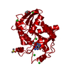
| Title | Crystal structure of Trichomonas vaginalis purine nucleoside phosphorylase | ||||||
 Components Components | purine nucleoside phosphorylase | ||||||
 Keywords Keywords | TRANSFERASE / alpha-beta-alpha sandwich | ||||||
| Function / homology | Nucleoside phosphorylase domain / Rossmann fold / 3-Layer(aba) Sandwich / Alpha Beta Function and homology information Function and homology information | ||||||
| Biological species |  Trichomonas vaginalis (eukaryote) Trichomonas vaginalis (eukaryote) | ||||||
| Method |  X-RAY DIFFRACTION / SYNCHROTRON / MOLECULAR REPLACEMENT / Resolution: 2.7 Å X-RAY DIFFRACTION / SYNCHROTRON / MOLECULAR REPLACEMENT / Resolution: 2.7 Å | ||||||
 Authors Authors | Zhang, Y. / Wang, W.H. / Wu, S.W. / Wang, C.C. / Ealick, S.E. | ||||||
 Citation Citation | Journal: J.Biol.Chem. / Year: 2005 Title: Identification of a subversive substrate of Trichomonas vaginalis purine nucleoside phosphorylase and the crystal structure of the enzyme-substrate complex. Authors: Zang, Y. / Wang, W.H. / Wu, S.W. / Ealick, S.E. / Wang, C.C. | ||||||
| History |
| ||||||
| Remark 999 | Sequence There is currently no match for the protein sequence in the standard databases |
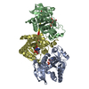
- Structure visualization
| Structure viewer | Molecule: MolmilJmol/JSmol |
|---|
- Downloads & links
-Download
| PDBx/mmCIF format | 1z33.cif.gz | 58.1 KB | Display | PDBx/mmCIF format |
|---|---|---|---|---|
| PDB format | pdb1z33.ent.gz | 42.6 KB | Display | PDB format |
| PDBx/mmJSON format | 1z33.json.gz | Tree view | PDBx/mmJSON format | |
| Others |  Other downloads Other downloads |
-Validation report
| Arichive directory | https://data.pdbj.org/pub/pdb/validation_reports/z3/1z33ftp://data.pdbj.org/pub/pdb/validation_reports/z3/1z33 | HTTPS FTP |
|---|
-Related structure data
| Related structure data | 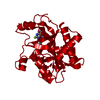 1z34C 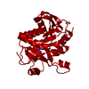 1z35C  1z36C 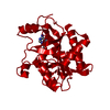 1z37C  1z38C  1z39C 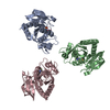 1pkeS C: citing same article ( S: Starting model for refinement |
|---|---|
| Similar structure data |
-Links
 PDBj
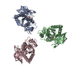
PDBj- Assembly
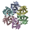
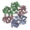
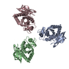
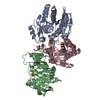
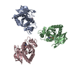
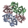
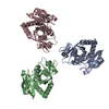
| Deposited unit | 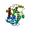
| |||||||||
|---|---|---|---|---|---|---|---|---|---|---|
| 1 | x 6
| |||||||||
| Unit cell |
| |||||||||
| Components on special symmetry positions |
|
-Components
| #1: Protein | Mass: 25813.658 Da / Num. of mol.: 1 Source method: isolated from a genetically manipulated source Source: (gene. exp.) Trichomonas vaginalis (eukaryote) / Production host:  |
|---|---|
| #2: Water | ChemComp-HOH /  Mass: 18.015 Da / Num. of mol.: 54 / Source method: isolated from a natural source / Formula: H2O Mass: 18.015 Da / Num. of mol.: 54 / Source method: isolated from a natural source / Formula: H2O |
-Experimental details
-Experiment
| Experiment | Method: X-RAY DIFFRACTION / Number of used crystals: 1 |
|---|
- Sample preparation
| Crystal | Density Matthews: 4.3 Å3/Da / Density % sol: 70 % |
|---|---|
| Crystal grow | Temperature: 291 K / Method: vapor diffusion, hanging drop / pH: 8.5 Details: PEG400, ethylene glycol, magnesium chloride, Tris, pH 8.5, VAPOR DIFFUSION, HANGING DROP, temperature 291K |
-Data collection
| Diffraction source | Source: SYNCHROTRON / Site: APS  / Beamline: 8-BM / Wavelength: 0.9795 / Wavelength: 0.9795 Å / Beamline: 8-BM / Wavelength: 0.9795 / Wavelength: 0.9795 Å |
|---|---|
| Radiation | Protocol: SINGLE WAVELENGTH / Monochromatic (M) / Laue (L): M / Scattering type: x-ray |
| Radiation wavelength | Wavelength: 0.9795 Å / Relative weight: 1 |
| Reflection | Resolution: 2.7→41.25 Å / Num. all: 12529 / Num. obs: 11760 / % possible obs: 93.6 % / Observed criterion σ(F): 0 / Observed criterion σ(I): 0 / Biso Wilson estimate: 34 Å2 |
| Reflection shell | Resolution: 2.7→2.87 Å / % possible all: 88.1 |
- Processing
| Software |
| ||||||||||||||||||||||||||||||||||||||||||||||||||||||||||||||||||||||||||||||||
|---|---|---|---|---|---|---|---|---|---|---|---|---|---|---|---|---|---|---|---|---|---|---|---|---|---|---|---|---|---|---|---|---|---|---|---|---|---|---|---|---|---|---|---|---|---|---|---|---|---|---|---|---|---|---|---|---|---|---|---|---|---|---|---|---|---|---|---|---|---|---|---|---|---|---|---|---|---|---|---|---|---|
| Refinement | Method to determine structure: MOLECULAR REPLACEMENT Starting model: 1PKE Resolution: 2.7→41.25 Å / Rfactor Rfree error: 0.011 / Data cutoff high absF: 270231.34 / Data cutoff low absF: 0 / Isotropic thermal model: RESTRAINED / Cross valid method: THROUGHOUT / σ(F): 0 / Stereochemistry target values: Engh & Huber
| ||||||||||||||||||||||||||||||||||||||||||||||||||||||||||||||||||||||||||||||||
| Solvent computation | Solvent model: FLAT MODEL / Bsol: 46.3946 Å2 / ksol: 0.372153 e/Å3 | ||||||||||||||||||||||||||||||||||||||||||||||||||||||||||||||||||||||||||||||||
| Displacement parameters | Biso mean: 48.5 Å2
| ||||||||||||||||||||||||||||||||||||||||||||||||||||||||||||||||||||||||||||||||
| Refine analyze |
| ||||||||||||||||||||||||||||||||||||||||||||||||||||||||||||||||||||||||||||||||
| Refinement step | Cycle: LAST / Resolution: 2.7→41.25 Å
| ||||||||||||||||||||||||||||||||||||||||||||||||||||||||||||||||||||||||||||||||
| Refine LS restraints |
| ||||||||||||||||||||||||||||||||||||||||||||||||||||||||||||||||||||||||||||||||
| LS refinement shell | Resolution: 2.7→2.87 Å / Rfactor Rfree error: 0.034 / Total num. of bins used: 6
| ||||||||||||||||||||||||||||||||||||||||||||||||||||||||||||||||||||||||||||||||
| Xplor file |
|
