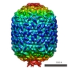
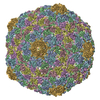
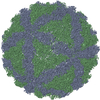
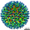
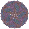
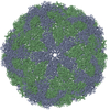
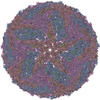
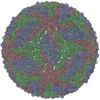
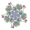
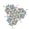
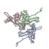
ジャーナル: Mol Cell / 年: 2005 タイトル: Conservation of the capsid structure in tailed dsDNA bacteriophages: the pseudoatomic structure of phi29. 著者: Marc C Morais / Kyung H Choi / Jaya S Koti / Paul R Chipman / Dwight L Anderson / Michael G Rossmann / 要旨: Bacteriophage phi29 is one of the smallest and simplest known dsDNA phages, making it amenable to structural investigations. The three-dimensional structure of a fiberless, isometric variant has been ...Bacteriophage phi29 is one of the smallest and simplest known dsDNA phages, making it amenable to structural investigations. The three-dimensional structure of a fiberless, isometric variant has been determined to 7.9 A resolution by cryo-electron microscopy (cryo-EM), allowing the identification of alpha helices and beta sheets. Their arrangement indicates that the folds of the phi29 and bacteriophage HK97 capsid proteins are similar except for an additional immunoglobulin-like domain of the phi29 protein. An atomic model that incorporates these two domains fits well into the cryo-EM density of the T = 3, fiberless isometric phi29 particle, and cryo-EM structures of fibered isometric and fiberless prolate prohead phi29 particles at resolutions of 8.7 A and 12.7 A, respectively. Thus, phi29 joins the growing number of phages that utilize the HK97 capsid structure, suggesting that this protein fold may be as prevalent in capsids of dsDNA phages as the jelly roll fold is in eukaryotic viruses.
SEQUENCE The authors state that an alignment between the coordinate C-alpha atoms and the sequence ...SEQUENCE The authors state that an alignment between the coordinate C-alpha atoms and the sequence is not possible. The following is the one-letter sequence for the protein modeled in this structure, SwissProt entry P13849: MRITFNDVKTSLGITESYDIVNAIRNSQGDNFKSYVPLATANNVAEVGAGILINQTVQND FITSLVDRIGLVVIRQVSLNNPLKKFKKGQIPLGRTIEEIYTDITKEKQYDAEEAEQKVF EREMPNVKTLFHERNRQGFYHQTIQDDSLKTAFVSWGNFESFVSSIINAIYNSAEVDEYE YMKLLVDNYYSKGLFTTVKIDEPTSSTGALTEFVKKMRATARKLTLPQGSRDWNSMAVRT RSYMEDLHLIIDADLEAELDVDVLAKAFNMNRTDFLGNVTVIDGFASTGLEAVLVDKDWF MVYDNLHKMETVRNPRGLYWNYYYHVWQTLSVSRFANAVAFVSGDVPAVTQVIVSPNIAA VKQGGQQQFTAYVRATNAKDHKVVWSVEGGSTGTAITGDGLLSVSGNEDNQLTVKATVDI GTEDKPKLVVGEAVVSIRPNNASGGAQA
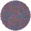
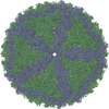
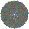
名称: Fiberless isometric phi29 capsid / タイプ: VIRUS 詳細: The capsid protein assembles a T=3 icosahedral virus shell. The virus particle is a protein shell with icosahedral symmetry. To generate the complete icosahedral particle, apply the following ...詳細: The capsid protein assembles a T=3 icosahedral virus shell. The virus particle is a protein shell with icosahedral symmetry. To generate the complete icosahedral particle, apply the following 59 matrices to the three chains in the icosahedral asymmetric unit, and combine with the original coordinates of the icosahedral asymmetric unit: 由来: NATURAL
由来(天然)
生物種: Enterobacteria phage T4 (ファージ)
ウイルスについての詳細
ホストのカテゴリ: BACTERIA / タイプ: VIRION
天然宿主
生物種: Bacillus subtilus
緩衝液
名称: tris-HCL / pH: 7.8 / 詳細: tris-HCL
試料
濃度: 1 mg/ml / 包埋: NO / シャドウイング: NO / 染色: NO / 凍結: YES
モード: BRIGHT FIELD / 倍率(公称値): 38000 X / 最大 デフォーカス(公称値): 3000 nm / 最小 デフォーカス(公称値): 700 nm / Cs: 2 mm
試料ホルダ
温度: 100 K / 傾斜角・最大: 0 ° / 傾斜角・最小: 0 °
撮影
電子線照射量: 20 e/Å2 / フィルム・検出器のモデル: KODAK SO-163 FILM
-
解析
EMソフトウェア
ID
名称
カテゴリ
詳細
1
EMfit
モデルフィッティング
2
SITUS COLORES
モデルフィッティング
3
SITUS COLORES
モデルフィッティング
4
EM3DR
3次元再構成
5
PFT
3次元再構成
EMPFT
6
POR
3次元再構成
CTF補正
詳細: Inverse of CTF was applied to images. Both phases and amplitudes were corrected.
対称性
点対称性: I (正20面体型対称)
3次元再構成
手法: 3D reconstructions were calculated using the Fourier-Bessel method. Initial orientations were found via common-lines, improved using model based polar Fourier transform methods, and finally ...手法: 3D reconstructions were calculated using the Fourier-Bessel method. Initial orientations were found via common-lines, improved using model based polar Fourier transform methods, and finally refined by minimizing the vector difference between between structure factors calculated from a particle image and those from a central section of the Fourier transform of the model. 解像度: 7.9 Å / 粒子像の数: 5922 / ピクセルサイズ(公称値): 1.8421 Å 詳細: Software used included P3DR (3D reconstructions), PFT (initial orientation improvement), and POR (final orientation refinement. 対称性のタイプ: POINT
原子モデル構築
プロトコル: RIGID BODY FIT / 空間: REAL Target criteria: rigid body refinement in real space against laplacian filtered EM density, using the program COLORES in the package SITUS. Each molecule in the T=3 asymmetric unit was refined separately. 詳細: METHOD--6D search for each symmetry related molecule in the icosahedral asymmetric unit REFINEMENT PROTOCOL--rigid body
 ムービー
ムービー コントローラー


 データを開く
 基本情報
基本情報 要素
要素 キーワード
キーワード
 Bacillus phage phi29 (ファージ)
 データ登録者
 引用
引用
 構造の表示
構造の表示 ムービービューア
 ダウンロードとリンク
 その他のダウンロード
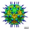


 PDBj
PDBj 集合体
集合体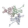


 試料調製
試料調製 電子顕微鏡撮影
 FIELD EMISSION GUN / 加速電圧: 200 kV / 照射モード: FLOOD BEAM
 解析
解析