 Movie
Movie Controller

[English] 日本語
 Yorodumi
- PDB-1ymv: SIGNAL TRANSDUCTION PROTEIN CHEY MUTANT WITH PHE 14 REPLACED BY G... -
+ Open data

- Basic information
| Entry | Database: PDB / ID: 1ymv | ||||||
|---|---|---|---|---|---|---|---|
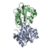
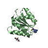
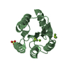
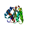
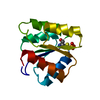
| Title | SIGNAL TRANSDUCTION PROTEIN CHEY MUTANT WITH PHE 14 REPLACED BY GLY, SER 15 REPLACED BY GLY, AND MET 17 REPLACED BY GLY | ||||||
 Components Components | CHEY | ||||||
 Keywords Keywords | CHEMOTAXIS / SENSORY TRANSDUCTION / PHOSPHORYLATION | ||||||
| Function / homology |  Function and homology information Function and homology informationbacterial-type flagellum basal body, C ring / bacterial-type flagellum rotor complex / bacterial-type flagellum-dependent swimming motility / aerotaxis / regulation of bacterial-type flagellum-dependent cell motility / thermotaxis / regulation of chemotaxis / internal peptidyl-lysine acetylation / bacterial-type flagellum / phosphorelay response regulator activity ...bacterial-type flagellum basal body, C ring / bacterial-type flagellum rotor complex / bacterial-type flagellum-dependent swimming motility / aerotaxis / regulation of bacterial-type flagellum-dependent cell motility / thermotaxis / regulation of chemotaxis / internal peptidyl-lysine acetylation / bacterial-type flagellum / phosphorelay response regulator activity / protein acetylation / acetyltransferase activity / phosphorelay signal transduction system / chemotaxis / magnesium ion binding / signal transduction / cytosol / cytoplasm Similarity search - Function | ||||||
| Biological species |  | ||||||
| Method |  X-RAY DIFFRACTION / Resolution: 1.9 Å X-RAY DIFFRACTION / Resolution: 1.9 Å | ||||||
 Authors Authors | Bellsolell, L. / Coll, M. | ||||||
 Citation Citation | Journal: J.Mol.Biol. / Year: 1996 Title: The three-dimensional structure of two mutants of the signal transduction protein CheY suggest its molecular activation mechanism. Authors: Bellsolell, L. / Cronet, P. / Majolero, M. / Serrano, L. / Coll, M. #1: Journal: J.Mol.Biol. / Year: 1995Title: Investigating the Structural Determinants of the P21-Like Triphosphate and Mg2+ Binding Site Authors: Cronet, P. / Bellsolell, L. / Sander, C. / Coll, M. / Serrano, L. #2: Journal: J.Mol.Biol. / Year: 1994Title: Magnesium Binding to the Bacterial Chemotaxis Protein Chey Results in Large Conformational Changes Involving its Functional Surface Authors: Bellsolell, L. / Prieto, J. / Serrano, L. / Coll, M. | ||||||
| History |
|
- Structure visualization
| Structure viewer | Molecule: MolmilJmol/JSmol |
|---|
- Downloads & links
-Download
| PDBx/mmCIF format | 1ymv.cif.gz | 38.2 KB | Display | PDBx/mmCIF format |
|---|---|---|---|---|
| PDB format | pdb1ymv.ent.gz | 25.8 KB | Display | PDB format |
| PDBx/mmJSON format | 1ymv.json.gz | Tree view | PDBx/mmJSON format | |
| Others |  Other downloads Other downloads |
-Validation report
| Arichive directory | https://data.pdbj.org/pub/pdb/validation_reports/ym/1ymvftp://data.pdbj.org/pub/pdb/validation_reports/ym/1ymv | HTTPS FTP |
|---|
-Related structure data


-Links
 PDBj
PDBj

- Assembly
| Deposited unit | 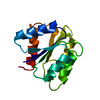
| ||||||||
|---|---|---|---|---|---|---|---|---|---|
| 1 |
| ||||||||
| Unit cell |
| ||||||||
| Atom site foot note | 1: CIS PROLINE - PRO 110 |
-Components
| #1: Protein | Mass: 13960.038 Da / Num. of mol.: 1 / Mutation: INS(M0), M1R, A2S, F14G, S15G, M17G Source method: isolated from a genetically manipulated source Details: MG2+ BOUND IN THE ACTIVE SITE / Source: (gene. exp.) |
|---|---|
| #2: Chemical | ChemComp-MG /   Mass: 24.305 Da / Num. of mol.: 1 / Source method: obtained synthetically / Formula: Mg Mass: 24.305 Da / Num. of mol.: 1 / Source method: obtained synthetically / Formula: Mg |
| #3: Water | ChemComp-HOH /  Mass: 18.015 Da / Num. of mol.: 87 / Source method: isolated from a natural source / Formula: H2O Mass: 18.015 Da / Num. of mol.: 87 / Source method: isolated from a natural source / Formula: H2O |
| Nonpolymer details | THE OCTAHEDRAL COORDINATION SHELL OF THE MG2+ ION INCLUDES CARBOXYL OXYGEN FROM ASP 13 AND ASP 57, ...THE OCTAHEDRAL |
-Experimental details
-Experiment
| Experiment | Method: X-RAY DIFFRACTION / Number of used crystals: 1 |
|---|
- Sample preparation
| Crystal | Density Matthews: 2.06 Å3/Da / Density % sol: 40.25 % | ||||||||||||||||||||||||||||||
|---|---|---|---|---|---|---|---|---|---|---|---|---|---|---|---|---|---|---|---|---|---|---|---|---|---|---|---|---|---|---|---|
| Crystal grow | pH: 6.8 / Details: pH 6.8 | ||||||||||||||||||||||||||||||
| Crystal grow | *PLUS Temperature: 4 ℃ / pH: 7.5 / Method: vapor diffusion, hanging drop | ||||||||||||||||||||||||||||||
| Components of the solutions | *PLUS
|
-Data collection
| Diffraction | Mean temperature: 277 K |
|---|---|
| Diffraction source | Wavelength: 1.5418 Å |
| Detector | Type: MARRESEARCH / Detector: IMAGE PLATE / Date: Dec 6, 1993 |
| Radiation | Monochromatic (M) / Laue (L): M / Scattering type: x-ray |
| Radiation wavelength | Wavelength: 1.5418 Å / Relative weight: 1 |
| Reflection | Resolution: 1.9→50 Å / Num. obs: 8100 / % possible obs: 84.9 % / Observed criterion σ(F): 2 / Rmerge(I) obs: 0.047 |
| Reflection | *PLUS Num. measured all: 27884 / Rmerge(I) obs: 0.047 |
| Reflection shell | *PLUS Highest resolution: 1.9 Å / Lowest resolution: 2 Å / % possible obs: 79 % |
- Processing
| Software |
| ||||||||||||||||||||||||||||||||||||||||||||||||||||||||||||
|---|---|---|---|---|---|---|---|---|---|---|---|---|---|---|---|---|---|---|---|---|---|---|---|---|---|---|---|---|---|---|---|---|---|---|---|---|---|---|---|---|---|---|---|---|---|---|---|---|---|---|---|---|---|---|---|---|---|---|---|---|---|
| Refinement | Resolution: 1.9→10 Å / σ(F): 2
| ||||||||||||||||||||||||||||||||||||||||||||||||||||||||||||
| Displacement parameters | Biso mean: 20 Å2 | ||||||||||||||||||||||||||||||||||||||||||||||||||||||||||||
| Refinement step | Cycle: LAST / Resolution: 1.9→10 Å
| ||||||||||||||||||||||||||||||||||||||||||||||||||||||||||||
| Refine LS restraints |
| ||||||||||||||||||||||||||||||||||||||||||||||||||||||||||||
| Software | *PLUS Name: X-PLOR / Classification: refinement | ||||||||||||||||||||||||||||||||||||||||||||||||||||||||||||
| Refinement | *PLUS | ||||||||||||||||||||||||||||||||||||||||||||||||||||||||||||
| Solvent computation | *PLUS | ||||||||||||||||||||||||||||||||||||||||||||||||||||||||||||
| Displacement parameters | *PLUS |
