 Movie
Movie Controller


+ Open data

- Basic information
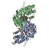
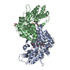
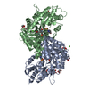
| Entry | Database: PDB / ID: 1yiy | ||||||
|---|---|---|---|---|---|---|---|
| Title | Aedes aegypti kynurenine aminotransferase | ||||||
 Components Components | kynurenine aminotransferase; glutamine transaminase K | ||||||
 Keywords Keywords | TRANSFERASE / kynurenine / kynurenic acid / kynurenine aminotrasferase / Aedes / mosquito / PLP-enzyme / PMP / pyridoxamine phosphate | ||||||
| Function / homology |  Function and homology information Function and homology information: / kynurenine-glyoxylate transaminase / L-kynurenine:glyoxylate transaminase activity / kynurenine-oxoglutarate transaminase / L-kynurenine:2-oxoglutarate transaminase activity / Transferases; Transferring nitrogenous groups; Transaminases / pyridoxal phosphate binding / mitochondrion Similarity search - Function | ||||||
| Biological species |  | ||||||
| Method |  X-RAY DIFFRACTION / SYNCHROTRON / MOLECULAR REPLACEMENT / Resolution: 1.9 Å X-RAY DIFFRACTION / SYNCHROTRON / MOLECULAR REPLACEMENT / Resolution: 1.9 Å | ||||||
 Authors Authors | Han, Q. / Gao, Y.G. / Robinson, H. / Ding, H. / Wilson, S. / Li, J. | ||||||
 Citation Citation | Journal: FEBS J. / Year: 2005 Title: Crystal structures of Aedes aegypti kynurenine aminotransferase. Authors: Han, Q. / Gao, Y.G. / Robinson, H. / Ding, H. / Wilson, S. / Li, J. | ||||||
| History |
|
- Structure visualization
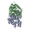
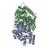
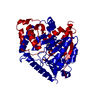
| Structure viewer | Molecule: MolmilJmol/JSmol |
|---|
- Downloads & links
-Download
| PDBx/mmCIF format | 1yiy.cif.gz | 184.5 KB | Display | PDBx/mmCIF format |
|---|---|---|---|---|
| PDB format | pdb1yiy.ent.gz | 145.7 KB | Display | PDB format |
| PDBx/mmJSON format | 1yiy.json.gz | Tree view | PDBx/mmJSON format | |
| Others |  Other downloads Other downloads |
-Validation report
| Arichive directory | https://data.pdbj.org/pub/pdb/validation_reports/yi/1yiyftp://data.pdbj.org/pub/pdb/validation_reports/yi/1yiy | HTTPS FTP |
|---|
-Related structure data
| Related structure data | 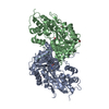 1yizC  1gckS  1v2dS C: citing same article ( S: Starting model for refinement |
|---|---|
| Similar structure data |


-Links
 PDBj
PDBj

- Assembly
| Deposited unit | 
| ||||||||
|---|---|---|---|---|---|---|---|---|---|
| 1 |
| ||||||||
| Unit cell |
|
-Components
| #1: Protein | Mass: 48336.137 Da / Num. of mol.: 2 / Fragment: kynurenine aminotransferase (Residues 49-477) Source method: isolated from a genetically manipulated source Source: (gene. exp.)  Spodoptera frugiperda (fall armyworm) / Strain (production host): sf9 Spodoptera frugiperda (fall armyworm) / Strain (production host): sf9References: UniProt: Q95VY4, UniProt: Q17CS8*PLUS, kynurenine-oxoglutarate transaminase, glutamine-phenylpyruvate transaminase #2: Chemical | ChemComp-BR /   Mass: 79.904 Da / Num. of mol.: 5 / Source method: obtained synthetically / Formula: Br Mass: 79.904 Da / Num. of mol.: 5 / Source method: obtained synthetically / Formula: Br#3: Chemical |   Mass: 248.173 Da / Num. of mol.: 2 / Source method: obtained synthetically / Formula: C8H13N2O5P Mass: 248.173 Da / Num. of mol.: 2 / Source method: obtained synthetically / Formula: C8H13N2O5P#4: Water | ChemComp-HOH / |  Mass: 18.015 Da / Num. of mol.: 440 / Source method: isolated from a natural source / Formula: H2O Mass: 18.015 Da / Num. of mol.: 440 / Source method: isolated from a natural source / Formula: H2O |
|---|
-Experimental details
-Experiment
| Experiment | Method: X-RAY DIFFRACTION / Number of used crystals: 1 |
|---|
- Sample preparation
| Crystal | Density Matthews: 2.29 Å3/Da / Density % sol: 46 % |
|---|---|
| Crystal grow | Temperature: 277 K / Method: vapor diffusion, hanging drop / pH: 8.5 Details: PEG 1000, TRIS.HCl, pH 8.5, VAPOR DIFFUSION, HANGING DROP, temperature 277K |
-Data collection
| Diffraction | Mean temperature: 123 K |
|---|---|
| Diffraction source | Source: SYNCHROTRON / Site: NSLS  / Beamline: X12C / Wavelength: 0.97 Å / Beamline: X12C / Wavelength: 0.97 Å |
| Detector | Type: SIEMENS FOUR-CIRCLE / Detector: DIFFRACTOMETER / Date: Aug 28, 2002 |
| Radiation | Protocol: SINGLE WAVELENGTH / Monochromatic (M) / Laue (L): M / Scattering type: x-ray |
| Radiation wavelength | Wavelength: 0.97 Å / Relative weight: 1 |
| Reflection | Resolution: 1.9→10 Å / Num. all: 68715 / Num. obs: 68715 / % possible obs: 97.1 % / Observed criterion σ(F): 0 / Observed criterion σ(I): 0 / Redundancy: 10.4 % / Rmerge(I) obs: 0.063 |
| Reflection shell | Resolution: 1.9→1.97 Å / % possible all: 90.5 |
- Processing
| Software |
| |||||||||||||||||||||||||
|---|---|---|---|---|---|---|---|---|---|---|---|---|---|---|---|---|---|---|---|---|---|---|---|---|---|---|
| Refinement | Method to determine structure: MOLECULAR REPLACEMENT Starting model: 1gck, 1v2d Resolution: 1.9→10 Å / Isotropic thermal model: Isotropic / Cross valid method: THROUGHOUT / σ(F): 4 / σ(I): 2 / Stereochemistry target values: Engh & Huber
| |||||||||||||||||||||||||
| Displacement parameters | Biso mean: 20.48 Å2 | |||||||||||||||||||||||||
| Refinement step | Cycle: LAST / Resolution: 1.9→10 Å
| |||||||||||||||||||||||||
| Refine LS restraints |
|
