 Movie
Movie Controller

[English] 日本語
 Yorodumi
- PDB-1ybv: STRUCTURE OF TRIHYDROXYNAPHTHALENE REDUCTASE IN COMPLEX WITH NADP... -
+ Open data

- Basic information
| Entry | Database: PDB / ID: 1ybv | ||||||
|---|---|---|---|---|---|---|---|
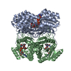
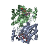
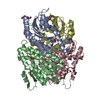
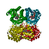
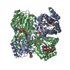
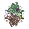
| Title | STRUCTURE OF TRIHYDROXYNAPHTHALENE REDUCTASE IN COMPLEX WITH NADPH AND AN ACTIVE SITE INHIBITOR | ||||||
 Components Components | TRIHYDROXYNAPHTHALENE REDUCTASE | ||||||
 Keywords Keywords | OXIDOREDUCTASE | ||||||
| Function / homology |  Function and homology information Function and homology informationtetrahydroxynaphthalene reductase / tetrahydroxynaphthalene reductase activity / melanin biosynthetic process Similarity search - Function | ||||||
| Biological species |  Magnaporthe grisea (fungus) Magnaporthe grisea (fungus) | ||||||
| Method |  X-RAY DIFFRACTION / Resolution: 2.8 Å X-RAY DIFFRACTION / Resolution: 2.8 Å | ||||||
 Authors Authors | Andersson, A. / Schneider, G. / Lindqvist, Y. | ||||||
 Citation Citation | Journal: Structure / Year: 1996 Title: Crystal structure of the ternary complex of 1,3,8-trihydroxynaphthalene reductase from Magnaporthe grisea with NADPH and an active-site inhibitor. Authors: Andersson, A. / Jordan, D. / Schneider, G. / Lindqvist, Y. #1: Journal: Proteins / Year: 1996Title: Crystallisation and Preliminary X-Ray Diffraction Study of 1,3,8-Trihydroxynaphthalene Reductase from Magnaporthe Grisea Authors: Andersson, A. / Jordan, D. / Schneider, G. / Valent, B. / Lindqvist, Y. | ||||||
| History |
|
- Structure visualization
| Structure viewer | Molecule: MolmilJmol/JSmol |
|---|
- Downloads & links
-Download
| PDBx/mmCIF format | 1ybv.cif.gz | 103.1 KB | Display | PDBx/mmCIF format |
|---|---|---|---|---|
| PDB format | pdb1ybv.ent.gz | 82 KB | Display | PDB format |
| PDBx/mmJSON format | 1ybv.json.gz | Tree view | PDBx/mmJSON format | |
| Others |  Other downloads Other downloads |
-Validation report
| Arichive directory | https://data.pdbj.org/pub/pdb/validation_reports/yb/1ybvftp://data.pdbj.org/pub/pdb/validation_reports/yb/1ybv | HTTPS FTP |
|---|
-Related structure data
| Similar structure data |
|---|


-Links
 PDBj
PDBj


- Assembly
| Deposited unit | 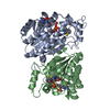
| ||||||||
|---|---|---|---|---|---|---|---|---|---|
| 1 | 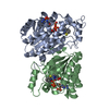
| ||||||||
| Unit cell |
| ||||||||
| Noncrystallographic symmetry (NCS) | NCS oper: (Code: given Matrix: (-0.999978, -0.004005, 0.005347), Vector: |
-Components
| #1: Protein | Mass: 30151.643 Da / Num. of mol.: 2 / Mutation: P2A, S241V, A242Q, H247 Source method: isolated from a genetically manipulated source Source: (gene. exp.) Magnaporthe grisea (fungus) / Strain: 409158 / Cell line: BL21 / Gene: BUF+ / Plasmid: PTHNR2 / Gene (production host): BUF+ / Production host:  #2: Chemical |   Mass: 745.421 Da / Num. of mol.: 2 / Source method: obtained synthetically / Formula: C21H30N7O17P3 Mass: 745.421 Da / Num. of mol.: 2 / Source method: obtained synthetically / Formula: C21H30N7O17P3#3: Chemical |   Mass: 190.245 Da / Num. of mol.: 2 / Source method: obtained synthetically / Formula: C9H8N3S Mass: 190.245 Da / Num. of mol.: 2 / Source method: obtained synthetically / Formula: C9H8N3S |
|---|
-Experimental details
-Experiment
| Experiment | Method: X-RAY DIFFRACTION |
|---|
- Sample preparation
| Crystal | Density Matthews: 3.55 Å3/Da / Density % sol: 58.5 % | ||||||||||||||||||||||||||||||||||||
|---|---|---|---|---|---|---|---|---|---|---|---|---|---|---|---|---|---|---|---|---|---|---|---|---|---|---|---|---|---|---|---|---|---|---|---|---|---|
| Crystal grow | *PLUS Method: vapor diffusion, hanging dropDetails: Andersson, A., (1996) Proteins: Struct.,Funct., Genet., 24, 525. | ||||||||||||||||||||||||||||||||||||
| Components of the solutions | *PLUS
|
-Data collection
| Diffraction source | Wavelength: 1.5418 |
|---|---|
| Detector | Type: RIGAKU RAXIS / Detector: IMAGE PLATE / Date: Dec 16, 1994 |
| Radiation | Monochromatic (M) / Laue (L): M / Scattering type: x-ray |
| Radiation wavelength | Wavelength: 1.5418 Å / Relative weight: 1 |
| Reflection | Num. obs: 19294 / % possible obs: 91.4 % / Observed criterion σ(I): 0 / Redundancy: 5.8 % / Rmerge(I) obs: 0.086 |
| Reflection | *PLUS Highest resolution: 2.8 Å / Num. measured all: 111124 |
- Processing
| Software |
| ||||||||||||||||||||||||||||||||||||||||||||||||||||||||||||
|---|---|---|---|---|---|---|---|---|---|---|---|---|---|---|---|---|---|---|---|---|---|---|---|---|---|---|---|---|---|---|---|---|---|---|---|---|---|---|---|---|---|---|---|---|---|---|---|---|---|---|---|---|---|---|---|---|---|---|---|---|---|
| Refinement | Resolution: 2.8→8 Å / σ(F): 2 / Details: STRICT TWO-FOLD NON-CRYSTALLOGRAPHIC SYMMETRY
| ||||||||||||||||||||||||||||||||||||||||||||||||||||||||||||
| Displacement parameters | Biso mean: 26.3 Å2 | ||||||||||||||||||||||||||||||||||||||||||||||||||||||||||||
| Refinement step | Cycle: LAST / Resolution: 2.8→8 Å
| ||||||||||||||||||||||||||||||||||||||||||||||||||||||||||||
| Refine LS restraints |
|
