HETEROGEN MN 353 IS AN INCOMPLETELY OCCUPIED MANGANESE, HOH 1002 IS MODELED TO ACCOUNT FOR RESIDUAL ...HETEROGEN MN 353 IS AN INCOMPLETELY OCCUPIED MANGANESE, HOH 1002 IS MODELED TO ACCOUNT FOR RESIDUAL NON-MANGANESE DENSITY AT THE NEARBY SPECIAL POSITION (TWO FOLD AXIS). NA 501 is intended to be on a special position (TWO FOLD AXIS).- it is about 0.26A away from its symmetry-related mate.
Remark 999
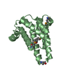
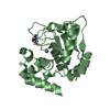
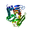
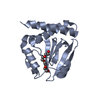
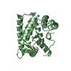
SEQUENCE the sequence is available in GeneDB (www.genedb.org) which is publicly available. The ...SEQUENCE the sequence is available in GeneDB (www.genedb.org) which is publicly available. The GeneDB identifier for this gene is LmjF30.1890. Additionally, residues 1-4 (GPGS-) are a cloning artifact; they are the remains of an N-terminal His tag. The remainder of the His tag was cleaved prior to crystallization. The Leishmanial protein consists of residues 5-184 noted in the SEQRES.
Resolution: 1.55→1.63 Å / Redundancy: 3.1 % / Mean I/σ(I) obs: 2 / Rsym value: 0.564 / % possible all: 94.8
-
Processing
Software
Name
Version
Classification
REFMAC
5.2.0005
refinement
MOSFLM
datareduction
CCP4
(SCALA)
datascaling
SHARP
phasing
Refinement
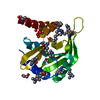
Method to determine structure: MAD / Resolution: 1.7→38 Å / Cor.coef. Fo:Fc: 0.957 / Cor.coef. Fo:Fc free: 0.938 / TLS residual ADP flag: LIKELY RESIDUAL / Isotropic thermal model: ISOTROPIC / Cross valid method: THROUGHOUT / σ(F): 0 / ESU R: 0.091 / ESU R Free: 0.091 / Stereochemistry target values: MA IMUM LIKELIHOOD Details: HYDROGENS HAVE BEEN ADDED IN THE RIDING POSITIONS. TWO LOOPS WHICH ARE PARTIALLY DISORDERED ARE MODELED WITH OCCUPANCY ESTIMATED AT 0.75 THESE INCLUDE RESIDUES 61-63 AND 120-123. SEVERAL ...Details: HYDROGENS HAVE BEEN ADDED IN THE RIDING POSITIONS. TWO LOOPS WHICH ARE PARTIALLY DISORDERED ARE MODELED WITH OCCUPANCY ESTIMATED AT 0.75 THESE INCLUDE RESIDUES 61-63 AND 120-123. SEVERAL DISORDERED SIDECHAIN ATOMS ARE MODELED WITH OCCUPANCY SET TO 0.10. PARTIALLY ORDERED SIDECHAIN ATOMS HAVE BEEN MODELED WITH OCCUPANCIES SET TO ONE-HALF OF THE CORRESPONDING MAIN CHAIN ATOMS. THE FINAL OCCUPANCIES OF THE PARTIALLY OCCUPIED HEAVY ATOMS WERE OBTAINED USING SHARP, AND ROUNDED TO THE NEAREST 0.05. THE CLOSE CONTACT BETWEEN RESIDUES GLU54 AND ILE63 IS BETWEEN SEVERELY DISORDERED SIDECHAINS IN A MINIMALLY ORDERED LOOP, AS NOTED above. THE OCCUPANCIES OF THESE SIDECHAINS HAS BEEN LOWERED TO REFLECT THE WEAK ELECTRON DENSITY IN THIS AREA OF THE MAPS.
Rfactor
Num. reflection
% reflection
Selection details
Rfree
0.19621
1181
5.1 %
RANDOM
Rwork
0.16492
-
-
-
all
0.1665
21985
-
-
obs
0.1665
21985
99.87 %
-
Solvent computation
Ion probe radii: 0.8 Å / Shrinkage radii: 0.8 Å / VDW probe radii: 1.2 Å / Solvent model: BABINET MODEL WITH MASK
Displacement parameters
Biso mean: 21.038 Å2
Baniso -1
Baniso -2
Baniso -3
1-
-0.16 Å2
0 Å2
0 Å2
2-
-
-0.16 Å2
0 Å2
3-
-
-
0.33 Å2
Refinement step
Cycle: LAST / Resolution: 1.7→38 Å
Protein
Nucleic acid
Ligand
Solvent
Total
Num. atoms
1366
0
42
157
1565
Refine LS restraints
Refine-ID
Type
Dev ideal
Dev ideal target
Number
X-RAY DIFFRACTION
r_bond_refined_d
0.011
0.021
1448
X-RAY DIFFRACTION
r_bond_other_d
0.001
0.02
1288
X-RAY DIFFRACTION
r_angle_refined_deg
1.467
1.991
1964
X-RAY DIFFRACTION
r_angle_other_deg
0.83
3
3012
X-RAY DIFFRACTION
r_dihedral_angle_1_deg
4.991
5
170
X-RAY DIFFRACTION
r_dihedral_angle_2_deg
31.532
24.933
75
X-RAY DIFFRACTION
r_dihedral_angle_3_deg
12.775
15
270
X-RAY DIFFRACTION
r_dihedral_angle_4_deg
16.196
15
10
X-RAY DIFFRACTION
r_chiral_restr
0.082
0.2
221
X-RAY DIFFRACTION
r_gen_planes_refined
0.006
0.02
1582
X-RAY DIFFRACTION
r_gen_planes_other
0.001
0.02
281
X-RAY DIFFRACTION
r_nbd_refined
0.226
0.2
298
X-RAY DIFFRACTION
r_nbd_other
0.188
0.2
1282
X-RAY DIFFRACTION
r_nbtor_refined
0.176
0.2
704
X-RAY DIFFRACTION
r_nbtor_other
0.082
0.2
778
X-RAY DIFFRACTION
r_metal_ion_refined
0.303
0.2
2
X-RAY DIFFRACTION
r_symmetry_vdw_refined
0.297
0.2
15
X-RAY DIFFRACTION
r_symmetry_vdw_other
0.298
0.2
40
X-RAY DIFFRACTION
r_symmetry_hbond_refined
0.216
0.2
12
X-RAY DIFFRACTION
r_mcbond_it
1.855
4
848
X-RAY DIFFRACTION
r_mcbond_other
0.584
4
345
X-RAY DIFFRACTION
r_mcangle_it
2.893
6
1381
X-RAY DIFFRACTION
r_scbond_it
3.369
6
615
X-RAY DIFFRACTION
r_scangle_it
5.249
10
583
LS refinement shell
Resolution: 1.7→1.792 Å / Total num. of bins used: 10
Rfactor
Num. reflection
% reflection
Rfree
0.228
152
-
Rwork
0.164
3166
-
obs
-
3318
99.94 %
Refinement TLS params.
Method: refined / Origin x: 27.726 Å / Origin y: 6.062 Å / Origin z: 17.465 Å
11
12
13
21
22
23
31
32
33
T
-0.0786 Å2
-0.0003 Å2
-0.0086 Å2
-
-0.0886 Å2
-0.0156 Å2
-
-
-0.0957 Å2
L
1.6244 °2
-0.4653 °2
0.1687 °2
-
1.1455 °2
-0.1666 °2
-
-
1.0981 °2
S
0.039 Å °
-0.0188 Å °
-0.0742 Å °
-0.0541 Å °
-0.028 Å °
0.0766 Å °
0.0314 Å °
0.0234 Å °
-0.011 Å °
+
About Yorodumi
-
News
-
Feb 9, 2022. New format data for meta-information of EMDB entries
New format data for meta-information of EMDB entries
Version 3 of the EMDB header file is now the official format.
The previous official version 1.9 will be removed from the archive.
In the structure databanks used in Yorodumi, some data are registered as the other names, "COVID-19 virus" and "2019-nCoV". Here are the details of the virus and the list of structure data.
Jan 31, 2019. EMDB accession codes are about to change! (news from PDBe EMDB page)
EMDB accession codes are about to change! (news from PDBe EMDB page)
The allocation of 4 digits for EMDB accession codes will soon come to an end. Whilst these codes will remain in use, new EMDB accession codes will include an additional digit and will expand incrementally as the available range of codes is exhausted. The current 4-digit format prefixed with “EMD-” (i.e. EMD-XXXX) will advance to a 5-digit format (i.e. EMD-XXXXX), and so on. It is currently estimated that the 4-digit codes will be depleted around Spring 2019, at which point the 5-digit format will come into force.
The EM Navigator/Yorodumi systems omit the EMD- prefix.
Related info.:Q: What is EMD? / ID/Accession-code notation in Yorodumi/EM Navigator
Yorodumi is a browser for structure data from EMDB, PDB, SASBDB, etc.
This page is also the successor to EM Navigator detail page, and also detail information page/front-end page for Omokage search.
The word "yorodu" (or yorozu) is an old Japanese word meaning "ten thousand". "mi" (miru) is to see.
Related info.:EMDB / PDB / SASBDB / Comparison of 3 databanks / Yorodumi Search / Aug 31, 2016. New EM Navigator & Yorodumi / Yorodumi Papers / Jmol/JSmol / Function and homology information / Changes in new EM Navigator and Yorodumi
 Movie
Movie Controller

 Yorodumi
 Open data
 Basic information
 Components
 Keywords
 Function and homology information
 Leishmania major (eukaryote)
 X-RAY DIFFRACTION /
  Authors
 Citation
 Structure visualization
 Downloads & links
 Other downloads


 PDBj
PDBj

 Assembly


 Mass: 54.938 Da / Num. of mol.: 8 / Source method: obtained synthetically / Formula: Mn
 Mass: 79.904 Da / Num. of mol.: 5 / Source method: obtained synthetically / Formula: Br
 Mass: 22.990 Da / Num. of mol.: 2 / Source method: obtained synthetically / Formula: Na
 Mass: 427.201 Da / Num. of mol.: 1 / Source method: obtained synthetically / Formula: C10H15N5O10P2 / Comment: ADP, energy-carrying molecule*YM
 Sample preparation
 / Beamline: 8.2.1 / Wavelength: 0.9198, 0.8856, 0.9202
 Processing