 Movie
Movie Controller

[English] 日本語
 Yorodumi
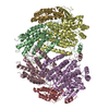
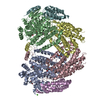
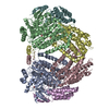
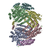
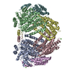
- PDB-1xvb: soluble methane monooxygenase hydroxylase: 6-bromohexanol soaked ... -
+ Open data

- Basic information
| Entry | Database: PDB / ID: 1xvb | ||||||
|---|---|---|---|---|---|---|---|
| Title | soluble methane monooxygenase hydroxylase: 6-bromohexanol soaked structure | ||||||
 Components Components | (Methane monooxygenase component A ...) x 3 | ||||||
 Keywords Keywords | OXIDOREDUCTASE / methane / diiron / cavities / products / four-helix bundle | ||||||
| Function / homology |  Function and homology information Function and homology informationmethane metabolic process / methane monooxygenase (soluble) / methane monooxygenase [NAD(P)H] activity / one-carbon metabolic process / metal ion binding Similarity search - Function | ||||||
| Biological species |  Methylococcus capsulatus (bacteria) Methylococcus capsulatus (bacteria) | ||||||
| Method |  X-RAY DIFFRACTION / SYNCHROTRON / MOLECULAR REPLACEMENT / Resolution: 1.8 Å X-RAY DIFFRACTION / SYNCHROTRON / MOLECULAR REPLACEMENT / Resolution: 1.8 Å | ||||||
 Authors Authors | Sazinsky, M.H. / Lippard, S.J. | ||||||
 Citation Citation | Journal: J.Am.Chem.Soc. / Year: 2005 Title: Product Bound Structures of the Soluble Methane Monooxygenase Hydroxylase from Methylococcus capsulatus (Bath): Protein Motion in the Alpha-Subunit Authors: Sazinsky, M.H. / Lippard, S.J. | ||||||
| History |
|
- Structure visualization
| Structure viewer | Molecule: MolmilJmol/JSmol |
|---|
- Downloads & links
-Download
| PDBx/mmCIF format | 1xvb.cif.gz | 459.9 KB | Display | PDBx/mmCIF format |
|---|---|---|---|---|
| PDB format | pdb1xvb.ent.gz | 370.5 KB | Display | PDB format |
| PDBx/mmJSON format | 1xvb.json.gz | Tree view | PDBx/mmJSON format | |
| Others |  Other downloads Other downloads |
-Validation report
| Arichive directory | https://data.pdbj.org/pub/pdb/validation_reports/xv/1xvbftp://data.pdbj.org/pub/pdb/validation_reports/xv/1xvb | HTTPS FTP |
|---|
-Related structure data
| Related structure data |  1xu3C  1xu5C  1xvcC  1xvdC  1xveC  1xvfC  1xvgC  1mtyS C: citing same article ( S: Starting model for refinement |
|---|---|
| Similar structure data |


-Links
 PDBj
PDBj
- Assembly
| Deposited unit | 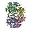
| ||||||||
|---|---|---|---|---|---|---|---|---|---|
| 1 |
| ||||||||
| Unit cell |
|
-Components
-Methane monooxygenase component A ... , 3 types, 6 molecules ABCDEF
| #1: Protein | Mass: 60719.113 Da / Num. of mol.: 2 / Source method: isolated from a natural source / Source: (natural) Methylococcus capsulatus (bacteria)References: UniProt: P22869, methane monooxygenase (soluble) #2: Protein | Mass: 45184.660 Da / Num. of mol.: 2 / Source method: isolated from a natural source / Source: (natural) Methylococcus capsulatus (bacteria)References: GenBank: 53804675, UniProt: P18798*PLUS, methane monooxygenase (soluble) #3: Protein | Mass: 19879.732 Da / Num. of mol.: 2 / Source method: isolated from a natural source / Source: (natural) Methylococcus capsulatus (bacteria)References: UniProt: P11987, methane monooxygenase (soluble) |
|---|
-Non-polymers , 7 types, 1135 molecules 
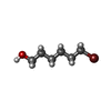
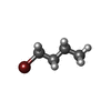
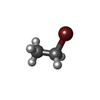

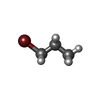

| #4: Chemical | ChemComp-FE /  Mass: 55.845 Da / Num. of mol.: 4 / Source method: obtained synthetically / Formula: Fe Mass: 55.845 Da / Num. of mol.: 4 / Source method: obtained synthetically / Formula: Fe#5: Chemical | ChemComp-BHL /  Mass: 181.071 Da / Num. of mol.: 10 / Source method: obtained synthetically / Formula: C6H13BrO Mass: 181.071 Da / Num. of mol.: 10 / Source method: obtained synthetically / Formula: C6H13BrO#6: Chemical | ChemComp-BBU / |  Mass: 137.018 Da / Num. of mol.: 1 / Source method: obtained synthetically / Formula: C4H9Br Mass: 137.018 Da / Num. of mol.: 1 / Source method: obtained synthetically / Formula: C4H9Br#7: Chemical | ChemComp-BBX / |  Mass: 108.965 Da / Num. of mol.: 1 / Source method: obtained synthetically / Formula: C2H5Br Mass: 108.965 Da / Num. of mol.: 1 / Source method: obtained synthetically / Formula: C2H5Br#8: Chemical | ChemComp-CA / |  Mass: 40.078 Da / Num. of mol.: 1 / Source method: obtained synthetically / Formula: Ca Mass: 40.078 Da / Num. of mol.: 1 / Source method: obtained synthetically / Formula: Ca#9: Chemical |  Mass: 122.992 Da / Num. of mol.: 2 / Source method: obtained synthetically / Formula: C3H7Br Mass: 122.992 Da / Num. of mol.: 2 / Source method: obtained synthetically / Formula: C3H7Br#10: Water | ChemComp-HOH / | Mass: 18.015 Da / Num. of mol.: 1116 / Source method: isolated from a natural source / Formula: H2O |
|---|
-Experimental details
-Experiment
| Experiment | Method: X-RAY DIFFRACTION / Number of used crystals: 1 |
|---|
- Sample preparation
| Crystal | Density Matthews: 2.3 Å3/Da / Density % sol: 55.1 % |
|---|---|
| Crystal grow | Temperature: 277 K / Method: vapor diffusion, hanging drop / pH: 7 Details: MOPS, PEG 8000, NaN3, CaCl2, glycerol, pH 7.0, VAPOR DIFFUSION, HANGING DROP, temperature 277K |
-Data collection
| Diffraction | Mean temperature: 100 K |
|---|---|
| Diffraction source | Source: SYNCHROTRON / Site: SSRL  / Beamline: BL11-1 / Wavelength: 1 Å / Beamline: BL11-1 / Wavelength: 1 Å |
| Detector | Type: ADSC QUANTUM 4 / Detector: CCD / Date: May 1, 2002 |
| Radiation | Monochromator: Si 111 Channel / Protocol: SINGLE WAVELENGTH / Monochromatic (M) / Laue (L): M / Scattering type: x-ray |
| Radiation wavelength | Wavelength: 1 Å / Relative weight: 1 |
| Reflection | Resolution: 1.8→30 Å / Num. all: 244201 / Num. obs: 244201 / % possible obs: 98.3 % / Observed criterion σ(F): 2 / Observed criterion σ(I): 2 / Redundancy: 3.9 % / Biso Wilson estimate: 21.5 Å2 / Rmerge(I) obs: 0.09 / Rsym value: 0.09 / Net I/σ(I): 16.8 |
| Reflection shell | Resolution: 1.8→1.88 Å / Rmerge(I) obs: 0.326 / Mean I/σ(I) obs: 2.8 / Rsym value: 0.326 / % possible all: 93.8 |
- Processing
| Software |
| ||||||||||||||||||||||||||||||||||||||||||||||||||||||||||||||||||||||||||||||||
|---|---|---|---|---|---|---|---|---|---|---|---|---|---|---|---|---|---|---|---|---|---|---|---|---|---|---|---|---|---|---|---|---|---|---|---|---|---|---|---|---|---|---|---|---|---|---|---|---|---|---|---|---|---|---|---|---|---|---|---|---|---|---|---|---|---|---|---|---|---|---|---|---|---|---|---|---|---|---|---|---|---|
| Refinement | Method to determine structure: MOLECULAR REPLACEMENT Starting model: 1mty Resolution: 1.8→29.85 Å / Rfactor Rfree error: 0.002 / Data cutoff high absF: 547689.5 / Data cutoff low absF: 0 / Isotropic thermal model: RESTRAINED / Cross valid method: THROUGHOUT / σ(F): 2 / σ(I): 2 / Stereochemistry target values: Engh & Huber
| ||||||||||||||||||||||||||||||||||||||||||||||||||||||||||||||||||||||||||||||||
| Solvent computation | Solvent model: FLAT MODEL / Bsol: 55.7582 Å2 / ksol: 0.36733 e/Å3 | ||||||||||||||||||||||||||||||||||||||||||||||||||||||||||||||||||||||||||||||||
| Displacement parameters | Biso mean: 37.1 Å2
| ||||||||||||||||||||||||||||||||||||||||||||||||||||||||||||||||||||||||||||||||
| Refine analyze |
| ||||||||||||||||||||||||||||||||||||||||||||||||||||||||||||||||||||||||||||||||
| Refinement step | Cycle: LAST / Resolution: 1.8→29.85 Å
| ||||||||||||||||||||||||||||||||||||||||||||||||||||||||||||||||||||||||||||||||
| Refine LS restraints |
| ||||||||||||||||||||||||||||||||||||||||||||||||||||||||||||||||||||||||||||||||
| LS refinement shell | Resolution: 1.8→1.91 Å / Rfactor Rfree error: 0.007 / Total num. of bins used: 6
| ||||||||||||||||||||||||||||||||||||||||||||||||||||||||||||||||||||||||||||||||
| Xplor file |
|
