 Movie
Movie Controller

[English] 日本語
 Yorodumi
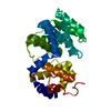
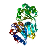
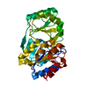
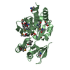
- PDB-1xqp: Crystal structure of 8-oxoguanosine complexed Pa-AGOG, 8-oxoguani... -
+ Open data

- Basic information
| Entry | Database: PDB / ID: 1xqp | ||||||
|---|---|---|---|---|---|---|---|
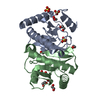
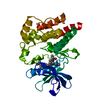
| Title | Crystal structure of 8-oxoguanosine complexed Pa-AGOG, 8-oxoguanine DNA glycosylase from Pyrobaculum aerophilum | ||||||
 Components Components | 8-oxoguanine DNA glycosylase | ||||||
 Keywords Keywords | LYASE / Helix-hairpin-Helix / 8-oxoguanine DNA glycosylase / Archaea / P.aerophilum / Pa-AGOG-8-oxoguanosine complex / DNA repair | ||||||
| Function / homology |  Function and homology information Function and homology informationoxidized base lesion DNA N-glycosylase activity / Hydrolases; Glycosylases; Hydrolysing N-glycosyl compounds / class I DNA-(apurinic or apyrimidinic site) endonuclease activity / DNA-(apurinic or apyrimidinic site) lyase / base-excision repair Similarity search - Function | ||||||
| Biological species |   Pyrobaculum aerophilum (archaea) Pyrobaculum aerophilum (archaea) | ||||||
| Method |  X-RAY DIFFRACTION / FOURIER SYNTHESIS / Resolution: 1.69 Å X-RAY DIFFRACTION / FOURIER SYNTHESIS / Resolution: 1.69 Å | ||||||
 Authors Authors | Lingaraju, G.M. / Sartori, A.A. / Kostrewa, D. / Prota, A.E. / Jiricny, J. / Winkler, F.K. | ||||||
 Citation Citation | Journal: Structure / Year: 2005 Title: A DNA glycosylase from Pyrobaculum aerophilum with an 8-oxoguanine binding mode and a noncanonical helix-hairpin-helix structure Authors: Lingaraju, G.M. / Sartori, A.A. / Kostrewa, D. / Prota, A.E. / Jiricny, J. / Winkler, F.K. #1: Journal: To be PublishedTitle: A novel highly thermostable 8-oxoguanine DNA glycosylase from Pyrobaculum aerophilum Authors: Sartori, A.A. / Lingaraju, G.M. / Hunziker, P. / Winkler, F.K. / Jiricny, J. | ||||||
| History |
|
- Structure visualization
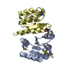
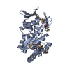
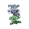
| Structure viewer | Molecule: MolmilJmol/JSmol |
|---|
- Downloads & links
-Download
| PDBx/mmCIF format | 1xqp.cif.gz | 70.1 KB | Display | PDBx/mmCIF format |
|---|---|---|---|---|
| PDB format | pdb1xqp.ent.gz | 51.2 KB | Display | PDB format |
| PDBx/mmJSON format | 1xqp.json.gz | Tree view | PDBx/mmJSON format | |
| Others |  Other downloads Other downloads |
-Validation report
| Arichive directory | https://data.pdbj.org/pub/pdb/validation_reports/xq/1xqpftp://data.pdbj.org/pub/pdb/validation_reports/xq/1xqp | HTTPS FTP |
|---|
-Related structure data

-Links
 PDBj
PDBj
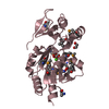
- Assembly
| Deposited unit | 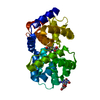
| ||||||||
|---|---|---|---|---|---|---|---|---|---|
| 1 |
| ||||||||
| Unit cell |
| ||||||||
| Components on special symmetry positions |
| ||||||||
| Details | The biologically active unit is a monomer. |
-Components
| #1: Protein | Mass: 29511.445 Da / Num. of mol.: 1 Source method: isolated from a genetically manipulated source Source: (gene. exp.) Pyrobaculum aerophilum (archaea) / Gene: Pa-AGOG / Plasmid: pET28C(+) / Production host:  | ||||
|---|---|---|---|---|---|
| #2: Chemical | 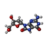  Mass: 283.241 Da / Num. of mol.: 2 / Source method: obtained synthetically / Formula: C10H13N5O5 Mass: 283.241 Da / Num. of mol.: 2 / Source method: obtained synthetically / Formula: C10H13N5O5#3: Water | ChemComp-HOH / |  Mass: 18.015 Da / Num. of mol.: 227 / Source method: isolated from a natural source / Formula: H2O Mass: 18.015 Da / Num. of mol.: 227 / Source method: isolated from a natural source / Formula: H2OHas protein modification | Y | |
-Experimental details
-Experiment
| Experiment | Method: X-RAY DIFFRACTION / Number of used crystals: 1 |
|---|
- Sample preparation
| Crystal | Density Matthews: 2.2 Å3/Da / Density % sol: 43 % |
|---|---|
| Crystal grow | Temperature: 295 K / Method: vapor diffusion, sitting drop / pH: 7.5 Details: PEG 4000, HEPES-NaOH, NaCl, Dithio threitol, Dimethyl sulfoxide, pH 7.5, VAPOR DIFFUSION, SITTING DROP, temperature 295K |
-Data collection
| Diffraction | Mean temperature: 100 K |
|---|---|
| Diffraction source | Source: ROTATING ANODE / Type: ENRAF-NONIUS FR591 / Wavelength: 1.5418 Å |
| Detector | Type: MAR scanner 345 mm plate / Detector: IMAGE PLATE / Date: Nov 14, 2003 / Details: Osmic mirrors |
| Radiation | Monochromator: Osmic Mirros / Protocol: SINGLE WAVELENGTH / Monochromatic (M) / Laue (L): M / Scattering type: x-ray |
| Radiation wavelength | Wavelength: 1.5418 Å / Relative weight: 1 |
| Reflection | Resolution: 1.69→57.74 Å / Num. all: 28623 / Num. obs: 28623 / % possible obs: 96.7 % / Observed criterion σ(F): 0 / Observed criterion σ(I): -3 / Redundancy: 6.8 % / Biso Wilson estimate: 32.6 Å2 / Rmerge(I) obs: 0.062 / Rsym value: 0.054 / Net I/σ(I): 17 |
| Reflection shell | Resolution: 1.69→1.8 Å / Redundancy: 6.12 % / Mean I/σ(I) obs: 3.47 / Num. unique all: 4548 / Rsym value: 0.578 / % possible all: 90.5 |
- Processing
| Software |
| ||||||||||||||||||||||||||||||||||||||||||||||||||||||||||||||||||||||||||||||||||||||||||||||||||||||||||||||||||||||||
|---|---|---|---|---|---|---|---|---|---|---|---|---|---|---|---|---|---|---|---|---|---|---|---|---|---|---|---|---|---|---|---|---|---|---|---|---|---|---|---|---|---|---|---|---|---|---|---|---|---|---|---|---|---|---|---|---|---|---|---|---|---|---|---|---|---|---|---|---|---|---|---|---|---|---|---|---|---|---|---|---|---|---|---|---|---|---|---|---|---|---|---|---|---|---|---|---|---|---|---|---|---|---|---|---|---|---|---|---|---|---|---|---|---|---|---|---|---|---|---|---|---|
| Refinement | Method to determine structure: FOURIER SYNTHESIS / Resolution: 1.69→40 Å / Cor.coef. Fo:Fc: 0.966 / Cor.coef. Fo:Fc free: 0.942 / SU B: 4.829 / SU ML: 0.084 / Isotropic thermal model: isotropic / Cross valid method: THROUGHOUT / σ(F): 0 / ESU R: 0.115 / ESU R Free: 0.114 / Stereochemistry target values: Engh & Huber / Details: HYDROGENS HAVE BEEN ADDED IN THE RIDING POSITIONS
| ||||||||||||||||||||||||||||||||||||||||||||||||||||||||||||||||||||||||||||||||||||||||||||||||||||||||||||||||||||||||
| Solvent computation | Ion probe radii: 0.8 Å / Shrinkage radii: 0.8 Å / VDW probe radii: 1.2 Å / Solvent model: MASK | ||||||||||||||||||||||||||||||||||||||||||||||||||||||||||||||||||||||||||||||||||||||||||||||||||||||||||||||||||||||||
| Displacement parameters | Biso mean: 28.831 Å2
| ||||||||||||||||||||||||||||||||||||||||||||||||||||||||||||||||||||||||||||||||||||||||||||||||||||||||||||||||||||||||
| Refine analyze |
| ||||||||||||||||||||||||||||||||||||||||||||||||||||||||||||||||||||||||||||||||||||||||||||||||||||||||||||||||||||||||
| Refinement step | Cycle: LAST / Resolution: 1.69→40 Å
| ||||||||||||||||||||||||||||||||||||||||||||||||||||||||||||||||||||||||||||||||||||||||||||||||||||||||||||||||||||||||
| Refine LS restraints |
| ||||||||||||||||||||||||||||||||||||||||||||||||||||||||||||||||||||||||||||||||||||||||||||||||||||||||||||||||||||||||
| LS refinement shell | Resolution: 1.69→1.734 Å / Total num. of bins used: 20 /
|
