 Movie
Movie Controller

[English] 日本語
 Yorodumi
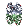
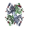
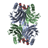
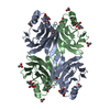
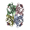
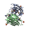
- PDB-1xi0: X-ray crystal structure of wild-type Xerocomus chrysenteron lectin XCL -
+ Open data

- Basic information
| Entry | Database: PDB / ID: 1xi0 | ||||||
|---|---|---|---|---|---|---|---|
| Title | X-ray crystal structure of wild-type Xerocomus chrysenteron lectin XCL | ||||||
 Components Components | lectin | ||||||
 Keywords Keywords | SUGAR BINDING PROTEIN / beta sandwich / greek key motif | ||||||
| Function / homology | Fungal fruit body lectin / Fungal fruit body lectin / Cytolysin/lectin / Cytolysin/lectin / Mutm (Fpg) Protein; Chain: A, domain 2 / Sandwich / Mainly Beta / Lectin Function and homology information Function and homology information | ||||||
| Biological species |  Xerocomus chrysenteron (red-cracking bolete) Xerocomus chrysenteron (red-cracking bolete) | ||||||
| Method |  X-RAY DIFFRACTION / SYNCHROTRON / MOLECULAR REPLACEMENT / Resolution: 2 Å X-RAY DIFFRACTION / SYNCHROTRON / MOLECULAR REPLACEMENT / Resolution: 2 Å | ||||||
 Authors Authors | Birck, C. / Damian, L. / Marty-Detraves, C. / Lougarre, A. / Schulze-Briese, C. / Koehl, P. / Fournier, D. / Paquereau, L. / Samama, J.P. | ||||||
 Citation Citation | Journal: J.Mol.Biol. / Year: 2004 Title: A new lectin family with structure similarity to actinoporins revealed by the crystal structure of Xerocomus chrysenteron lectin XCL Authors: Birck, C. / Damian, L. / Marty-Detraves, C. / Lougarre, A. / Schulze-Briese, C. / Koehl, P. / Fournier, D. / Paquereau, L. / Samama, J.P. | ||||||
| History |
|
- Structure visualization
| Structure viewer | Molecule: MolmilJmol/JSmol |
|---|
- Downloads & links
-Download
| PDBx/mmCIF format | 1xi0.cif.gz | 65.2 KB | Display | PDBx/mmCIF format |
|---|---|---|---|---|
| PDB format | pdb1xi0.ent.gz | 48.4 KB | Display | PDB format |
| PDBx/mmJSON format | 1xi0.json.gz | Tree view | PDBx/mmJSON format | |
| Others |  Other downloads Other downloads |
-Validation report
| Summary document | 1xi0_validation.pdf.gz | 433.9 KB | Display | wwPDB validaton report |
|---|---|---|---|---|
| Full document | 1xi0_full_validation.pdf.gz | 438.5 KB | Display | |
| Data in XML | 1xi0_validation.xml.gz | 13.3 KB | Display | |
| Data in CIF | 1xi0_validation.cif.gz | 17.4 KB | Display | |
| Arichive directory | https://data.pdbj.org/pub/pdb/validation_reports/xi/1xi0ftp://data.pdbj.org/pub/pdb/validation_reports/xi/1xi0 | HTTPS FTP |
-Related structure data
| Related structure data |  1x99SC S: Starting model for refinement C: citing same article ( |
|---|---|
| Similar structure data |


-Links
 PDBj
PDBj- Assembly
| Deposited unit | 
| ||||||||
|---|---|---|---|---|---|---|---|---|---|
| 1 | 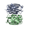
| ||||||||
| Unit cell |
| ||||||||
| Components on special symmetry positions |
| ||||||||
| Details | the biological assembly is a tetramer generated from the dimer in the asymetric unit by the operation: -x, -y, z |
-Components
| #1: Protein | Mass: 16031.808 Da / Num. of mol.: 2 Source method: isolated from a genetically manipulated source Source: (gene. exp.) Xerocomus chrysenteron (red-cracking bolete)Plasmid: modified pET28 / Species (production host): Escherichia coli / Production host:  #2: Water | ChemComp-HOH / |  Mass: 18.015 Da / Num. of mol.: 65 / Source method: isolated from a natural source / Formula: H2O Mass: 18.015 Da / Num. of mol.: 65 / Source method: isolated from a natural source / Formula: H2O |
|---|
-Experimental details
-Experiment
| Experiment | Method: X-RAY DIFFRACTION / Number of used crystals: 1 |
|---|
- Sample preparation
| Crystal | Density Matthews: 2.3 Å3/Da / Density % sol: 45.5 % |
|---|---|
| Crystal grow | Temperature: 295 K / Method: vapor diffusion, hanging drop / pH: 6.8 Details: ammonium sulfate, MES, zinc chloride, pH 6.8, VAPOR DIFFUSION, HANGING DROP, temperature 295K |
-Data collection
| Diffraction | Mean temperature: 100 K |
|---|---|
| Diffraction source | Source: SYNCHROTRON / Site: ESRF  / Beamline: ID14-1 / Wavelength: 0.934 Å / Beamline: ID14-1 / Wavelength: 0.934 Å |
| Detector | Type: ADSC QUANTUM 4 / Detector: CCD / Date: Sep 14, 2002 |
| Radiation | Monochromator: Diamond (111), Ge (220) / Protocol: SINGLE WAVELENGTH / Monochromatic (M) / Laue (L): M / Scattering type: x-ray |
| Radiation wavelength | Wavelength: 0.934 Å / Relative weight: 1 |
| Reflection | Resolution: 1.98→50 Å / Num. all: 89122 / Num. obs: 20047 / % possible obs: 99.6 % / Observed criterion σ(F): 2 / Observed criterion σ(I): 2 / Redundancy: 4.4 % / Biso Wilson estimate: 27.9 Å2 / Rsym value: 0.062 / Net I/σ(I): 17.3 |
| Reflection shell | Resolution: 1.98→2.05 Å / Redundancy: 3.8 % / Mean I/σ(I) obs: 6.2 / Num. unique all: 2010 / Rsym value: 0.272 / % possible all: 99.1 |
- Processing
| Software |
| |||||||||||||||||||||||||
|---|---|---|---|---|---|---|---|---|---|---|---|---|---|---|---|---|---|---|---|---|---|---|---|---|---|---|
| Refinement | Method to determine structure: MOLECULAR REPLACEMENT Starting model: pdb entry 1x99 Resolution: 2→50 Å / Isotropic thermal model: anisotropic / σ(F): 0 / Stereochemistry target values: Engh & Huber
| |||||||||||||||||||||||||
| Refine analyze | Luzzati coordinate error obs: 0.27 Å / Luzzati d res low obs: 5 Å / Luzzati sigma a obs: 0.32 Å | |||||||||||||||||||||||||
| Refinement step | Cycle: LAST / Resolution: 2→50 Å
| |||||||||||||||||||||||||
| Refine LS restraints |
| |||||||||||||||||||||||||
| LS refinement shell | Resolution: 2→2.07 Å / Rfactor Rfree error: 0.009
|
