 Movie
Movie Controller

[English] 日本語
 Yorodumi
- PDB-1xfs: X-Ray Crystal Structure of Protein NE0264 from Nitrosomonas europ... -
+ Open data

- Basic information
| Entry | Database: PDB / ID: 1xfs | ||||||
|---|---|---|---|---|---|---|---|
| Title | X-Ray Crystal Structure of Protein NE0264 from Nitrosomonas europaea. Northeast Structural Genomics Consortium Target NeR5. | ||||||
 Components Components | conserved hypothetical protein | ||||||
 Keywords Keywords | STRUCTURAL GENOMICS / UNKNOWN FUNCTION / Protein Structure Initiative / NESG / alpha-beta protein / PSI / Northeast Structural Genomics Consortium | ||||||
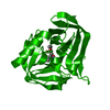
| Function / homology | Activator of Hsp90 ATPase homologue 1-like / Activator of Hsp90 ATPase homolog 1-like protein / START domain / Alpha-D-Glucose-1,6-Bisphosphate; Chain A, domain 4 / START-like domain superfamily / 2-Layer Sandwich / Alpha Beta / Activator of Hsp90 ATPase homologue 1/2-like C-terminal domain-containing protein Function and homology information Function and homology information | ||||||
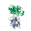
| Biological species |  Nitrosomonas europaea (bacteria) Nitrosomonas europaea (bacteria) | ||||||
| Method |  X-RAY DIFFRACTION / SYNCHROTRON / SAD / Resolution: 1.7 Å X-RAY DIFFRACTION / SYNCHROTRON / SAD / Resolution: 1.7 Å | ||||||
 Authors Authors | Forouhar, F. / Abashidze, M. / Vorobiev, S.M. / Ciano, M. / Ma, L.-C. / Shastry, R. / Acton, T.B. / Montelione, G.T. / Tong, L. / Hunt, J.F. / Northeast Structural Genomics Consortium (NESG) | ||||||
 Citation Citation | Journal: To be Published Title: Crystal Structure of the hypothetical Protein from Nitrosomonas europaea, NESG Target NeR5 Authors: Forouhar, F. / Abashidze, M. / Vorobiev, S.M. / Ciano, M. / Ma, L.-C. / Shastry, R. / Acton, T.B. / Montelione, G.T. / Tong, L. / Hunt, J.F. | ||||||
| History |
|
- Structure visualization
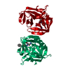
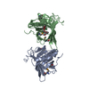
| Structure viewer | Molecule: MolmilJmol/JSmol |
|---|
- Downloads & links
-Download
| PDBx/mmCIF format | 1xfs.cif.gz | 82.4 KB | Display | PDBx/mmCIF format |
|---|---|---|---|---|
| PDB format | pdb1xfs.ent.gz | 61.4 KB | Display | PDB format |
| PDBx/mmJSON format | 1xfs.json.gz | Tree view | PDBx/mmJSON format | |
| Others |  Other downloads Other downloads |
-Validation report
| Summary document | 1xfs_validation.pdf.gz | 441 KB | Display | wwPDB validaton report |
|---|---|---|---|---|
| Full document | 1xfs_full_validation.pdf.gz | 447.6 KB | Display | |
| Data in XML | 1xfs_validation.xml.gz | 20.1 KB | Display | |
| Data in CIF | 1xfs_validation.cif.gz | 28 KB | Display | |
| Arichive directory | https://data.pdbj.org/pub/pdb/validation_reports/xf/1xfsftp://data.pdbj.org/pub/pdb/validation_reports/xf/1xfs | HTTPS FTP |
-Related structure data
| Similar structure data | |
|---|---|
| Other databases |


-Links
 PDBj
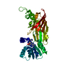
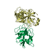
PDBj- Assembly
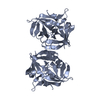
| Deposited unit | 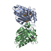
| ||||||||
|---|---|---|---|---|---|---|---|---|---|
| 1 |
| ||||||||
| Unit cell |
|
-Components
| #1: Protein | Mass: 20610.746 Da / Num. of mol.: 2 Source method: isolated from a genetically manipulated source Source: (gene. exp.) Nitrosomonas europaea (bacteria) / Strain: ATCC 19718 / Plasmid: BL21 / Production host: #2: Water | ChemComp-HOH / |  Mass: 18.015 Da / Num. of mol.: 342 / Source method: isolated from a natural source / Formula: H2O Mass: 18.015 Da / Num. of mol.: 342 / Source method: isolated from a natural source / Formula: H2OHas protein modification | Y | |
|---|
-Experimental details
-Experiment
| Experiment | Method: X-RAY DIFFRACTION / Number of used crystals: 1 |
|---|
- Sample preparation
| Crystal | Density Matthews: 2.55 Å3/Da / Density % sol: 50 % |
|---|---|
| Crystal grow | Temperature: 293 K / Method: vapor diffusion, hanging drop / pH: 6.5 Details: 100mM Cacodylate Acid (pH 6.5), 20%PEG3350, 200mM ammonium Citrate, VAPOR DIFFUSION, HANGING DROP, temperature 293K |
-Data collection
| Diffraction | Mean temperature: 100 K |
|---|---|
| Diffraction source | Source: SYNCHROTRON / Site: NSLS  / Beamline: X4A / Wavelength: 0.97902 Å / Beamline: X4A / Wavelength: 0.97902 Å |
| Detector | Type: ADSC QUANTUM 4 / Detector: CCD / Date: Aug 5, 2004 / Details: mirrors |
| Radiation | Monochromator: Si 111 CHANNEL / Protocol: SINGLE WAVELENGTH / Monochromatic (M) / Laue (L): M / Scattering type: x-ray |
| Radiation wavelength | Wavelength: 0.97902 Å / Relative weight: 1 |
| Reflection | Resolution: 1.7→27.79 Å / Num. all: 287876 / Num. obs: 71306 / % possible obs: 93.3 % / Observed criterion σ(F): 2 / Observed criterion σ(I): 2 / Redundancy: 4 % / Biso Wilson estimate: 16 Å2 / Rmerge(I) obs: 0.033 / Rsym value: 0.036 / Net I/σ(I): 41.6 |
| Reflection shell | Resolution: 1.7→1.76 Å / Redundancy: 3.8 % / Rmerge(I) obs: 0.078 / Mean I/σ(I) obs: 14.8 / Num. unique all: 7100 / Rsym value: 0.074 / % possible all: 92.5 |
- Processing
| Software |
| |||||||||||||||||||||||||
|---|---|---|---|---|---|---|---|---|---|---|---|---|---|---|---|---|---|---|---|---|---|---|---|---|---|---|
| Refinement | Method to determine structure: SAD / Resolution: 1.7→27.79 Å / Rfactor Rfree error: 0.003 / Data cutoff high absF: 1041734.09 / Data cutoff low absF: 0 / Cross valid method: THROUGHOUT / σ(F): 2 / σ(I): 2 / Stereochemistry target values: Engh & Huber
| |||||||||||||||||||||||||
| Solvent computation | Solvent model: FLAT MODEL / Bsol: 56.1999 Å2 / ksol: 0.387838 e/Å3 | |||||||||||||||||||||||||
| Displacement parameters | Biso mean: 21.4 Å2
| |||||||||||||||||||||||||
| Refine analyze |
| |||||||||||||||||||||||||
| Refinement step | Cycle: LAST / Resolution: 1.7→27.79 Å
| |||||||||||||||||||||||||
| Refine LS restraints |
| |||||||||||||||||||||||||
| LS refinement shell | Resolution: 1.7→1.81 Å / Rfactor Rfree error: 0.009 / Total num. of bins used: 6
|
