- PDB-1xea: Crystal structure of a Gfo/Idh/MocA family oxidoreductase from Vi... -
+
Open data
ID or keywords:
Loading...
-
Basic information
Entry
Database: PDB / ID: 1xea
Title
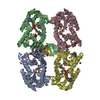
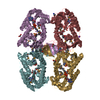
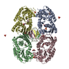
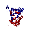
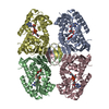
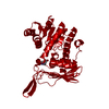
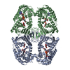
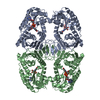
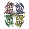
Crystal structure of a Gfo/Idh/MocA family oxidoreductase from Vibrio cholerae
Components
Oxidoreductase, Gfo/Idh/MocA family
Keywords
OXIDOREDUCTASE / Structural Genomics / Protein Structure Initiative / NYSGXRC / T1536 / VCA1048 / Gfo/Idh/MocA family oxidoreductase / PSI / New York SGX Research Center for Structural Genomics
A: Oxidoreductase, Gfo/Idh/MocA family B: Oxidoreductase, Gfo/Idh/MocA family C: Oxidoreductase, Gfo/Idh/MocA family D: Oxidoreductase, Gfo/Idh/MocA family hetero molecules
Resolution: 2.65→2.74 Å / Mean I/σ(I) obs: 1.3 / Num. unique all: 8472 / Rsym value: 0.61 / % possible all: 92.4
-
Processing
Software
Name
Version
Classification
DENZO
datareduction
SCALEPACK
datascaling
MLPHARE
phasing
CNS
1.1
refinement
Refinement
Method to determine structure: MOLECULAR REPLACEMENT, SAD Starting model: Experimental electron density map from Se-SAD phasing. Resolution: 2.65→19.99 Å / Rfactor Rfree error: 0.004 / Data cutoff high absF: 180192.73 / Data cutoff low absF: 0 / Isotropic thermal model: RESTRAINED / Cross valid method: THROUGHOUT / σ(F): 0 Details: A poor molecular replacement solution was obtained using a model derived from pdb entry 1TLT. MR phases were used to locate Selenium sites. One unknown metal ion was located. It was refined ...Details: A poor molecular replacement solution was obtained using a model derived from pdb entry 1TLT. MR phases were used to locate Selenium sites. One unknown metal ion was located. It was refined as partially occupied Ni+2 (may be from purification column).
In the structure databanks used in Yorodumi, some data are registered as the other names, "COVID-19 virus" and "2019-nCoV". Here are the details of the virus and the list of structure data.
Jan 31, 2019. EMDB accession codes are about to change! (news from PDBe EMDB page)
EMDB accession codes are about to change! (news from PDBe EMDB page)
The allocation of 4 digits for EMDB accession codes will soon come to an end. Whilst these codes will remain in use, new EMDB accession codes will include an additional digit and will expand incrementally as the available range of codes is exhausted. The current 4-digit format prefixed with “EMD-” (i.e. EMD-XXXX) will advance to a 5-digit format (i.e. EMD-XXXXX), and so on. It is currently estimated that the 4-digit codes will be depleted around Spring 2019, at which point the 5-digit format will come into force.
The EM Navigator/Yorodumi systems omit the EMD- prefix.
Related info.:Q: What is EMD? / ID/Accession-code notation in Yorodumi/EM Navigator
Yorodumi is a browser for structure data from EMDB, PDB, SASBDB, etc.
This page is also the successor to EM Navigator detail page, and also detail information page/front-end page for Omokage search.
The word "yorodu" (or yorozu) is an old Japanese word meaning "ten thousand". "mi" (miru) is to see.
Related info.:EMDB / PDB / SASBDB / Comparison of 3 databanks / Yorodumi Search / Aug 31, 2016. New EM Navigator & Yorodumi / Yorodumi Papers / Jmol/JSmol / Function and homology information / Changes in new EM Navigator and Yorodumi
 Movie
Movie Controller

 Yorodumi
 Open data
 Basic information
 Components
 Keywords
 Function and homology information
 Vibrio cholerae (bacteria)
 X-RAY DIFFRACTION /
  Authors
 Citation
 Structure visualization
 Downloads & links
 Other downloads
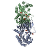
 PDBj
PDBj Assembly
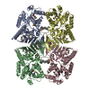

 Mass: 58.693 Da / Num. of mol.: 1 / Source method: obtained synthetically / Formula: Ni
 Mass: 18.015 Da / Num. of mol.: 273 / Source method: isolated from a natural source / Formula: H2O
 Sample preparation
 / Beamline: 31-ID / Wavelength: 0.98 Å
 Processing