- PDB-3cea: Crystal structure of myo-inositol 2-dehydrogenase (NP_786804.1) f... -
+
Open data
ID or keywords:
Loading...
-
Basic information
Entry
Database: PDB / ID: 3cea
Title
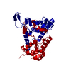
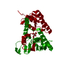
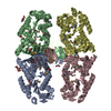
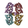
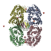
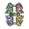
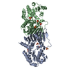
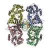
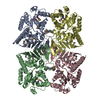
Crystal structure of myo-inositol 2-dehydrogenase (NP_786804.1) from Lactobacillus plantarum at 2.40 A resolution
Components
Myo-inositol 2-dehydrogenase
Keywords
OXIDOREDUCTASE / NP_786804.1 / myo-inositol 2-dehydrogenase / Oxidoreductase family / NAD-binding Rossmann fold / Structural Genomics / Joint Center for Structural Genomics / JCSG / Protein Structure Initiative / PSI-2
Function / homology
Function and homology information
NADPH regeneration / Oxidoreductases; Acting on the CH-OH group of donors; With NAD+ or NADP+ as acceptor / oxidoreductase activity / nucleotide binding / cytoplasm Similarity search - Function
Mass: 18.015 Da / Num. of mol.: 343 / Source method: isolated from a natural source / Formula: H2O
Has protein modification
Y
Sequence details
THE CONSTRUCT WAS EXPRESSED WITH A PURIFICATION TAG MGSDKIHHHHHHENLYFQG. THE TAG WAS REMOVED WITH ...THE CONSTRUCT WAS EXPRESSED WITH A PURIFICATION TAG MGSDKIHHHHHHENLYFQG. THE TAG WAS REMOVED WITH TEV PROTEASE LEAVING ONLY A GLYCINE (0) FOLLOWED BY THE TARGET SEQUENCE.
-
Experimental details
-
Experiment
Experiment
Method: X-RAY DIFFRACTION / Number of used crystals: 1
-
Sample preparation
Crystal
Density Matthews: 2.23 Å3/Da / Density % sol: 44.84 %
Crystal grow
Temperature: 277 K / Method: vapor diffusion, sitting drop / pH: 6.5 Details: NANODROP, 0.2M MgCl2, 20.0% PEG 1000, 0.1M Cacodylate pH 6.5, VAPOR DIFFUSION, SITTING DROP, temperature 277K
Type: MARMOSAIC 325 mm CCD / Detector: CCD / Date: Dec 10, 2007 / Details: Flat mirror (vertical focusing)
Radiation
Monochromator: Single crystal Si(111) bent (horizontal focusing) Protocol: MAD / Monochromatic (M) / Laue (L): M / Scattering type: x-ray
Radiation wavelength
ID
Wavelength (Å)
Relative weight
1
0.91837
1
2
0.97941
1
3
0.97883
1
Reflection
Resolution: 2.4→29.45 Å / Num. obs: 52411 / % possible obs: 99 % / Redundancy: 3.6 % / Biso Wilson estimate: 33.924 Å2 / Rmerge(I) obs: 0.133 / Rsym value: 0.133 / Net I/σ(I): 9.3
Reflection shell
Diffraction-ID: 1
Resolution (Å)
Redundancy (%)
Rmerge(I) obs
Mean I/σ(I) obs
Num. measured all
Num. unique all
Rsym value
% possible all
2.4-2.46
2
0.429
1.9
7021
3467
0.429
91.8
2.46-2.53
2
0.388
1.9
7315
3575
0.388
94.5
2.53-2.6
3.8
0.517
1.4
14404
3746
0.517
100
2.6-2.68
3.9
0.447
1.6
13844
3592
0.447
100
2.68-2.77
3.8
0.364
2
13370
3484
0.364
100
2.77-2.87
3.9
0.293
2.5
13082
3383
0.293
100
2.87-2.98
3.9
0.265
2.8
12644
3283
0.265
100
2.98-3.1
3.9
0.217
3.4
12073
3124
0.217
100
3.1-3.24
3.9
0.176
4.2
11606
3005
0.176
100
3.24-3.39
3.9
0.139
5.3
11311
2935
0.139
100
3.39-3.58
3.9
0.111
6.5
10477
2712
0.111
100
3.58-3.79
3.9
0.092
7.8
9951
2577
0.092
100
3.79-4.06
3.9
0.083
8.6
9457
2453
0.083
100
4.06-4.38
3.8
0.077
8.8
8804
2290
0.077
100
4.38-4.8
3.8
0.072
8.9
8091
2104
0.072
100
4.8-5.37
3.9
0.072
9.6
7272
1888
0.072
100
5.37-6.2
3.9
0.08
9
6452
1670
0.08
100
6.2-7.59
3.8
0.069
10.3
5442
1422
0.069
100
7.59-10.73
3.8
0.048
13.4
4210
1106
0.048
100
10.73-29.45
3.6
0.045
13.5
2171
595
0.045
95.1
-
Phasing
Phasing
Method: MAD
-
Processing
Software
Name
Version
Classification
NB
REFMAC
5.2.0019
refinement
PHENIX
refinement
SHELX
phasing
MolProbity
3beta29
modelbuilding
SCALA
datascaling
PDB_EXTRACT
3
dataextraction
MAR345
CCD
datacollection
MOSFLM
datareduction
SHELXD
phasing
SHARP
phasing
Refinement
Method to determine structure: MAD / Resolution: 2.4→29.45 Å / Cor.coef. Fo:Fc: 0.942 / Cor.coef. Fo:Fc free: 0.909 / SU B: 19.09 / SU ML: 0.224 / TLS residual ADP flag: LIKELY RESIDUAL / Cross valid method: THROUGHOUT / σ(F): 0 / ESU R: 0.655 / ESU R Free: 0.28 Stereochemistry target values: MAXIMUM LIKELIHOOD WITH PHASES Details: 1. HYDROGENS HAVE BEEN ADDED IN THE RIDING POSITIONS. 2. A MET-INHIBITION PROTOCOL WAS USED FOR SELENOMETHIONINE INCORPORATION DURING PROTEIN EXPRESSION. THE OCCUPANCY OF THE SE ATOMS IN THE ...Details: 1. HYDROGENS HAVE BEEN ADDED IN THE RIDING POSITIONS. 2. A MET-INHIBITION PROTOCOL WAS USED FOR SELENOMETHIONINE INCORPORATION DURING PROTEIN EXPRESSION. THE OCCUPANCY OF THE SE ATOMS IN THE MSE RESIDUES WAS REDUCED TO 0.75 FOR THE REDUCED SCATTERING POWER DUE TO PARTIAL S-MET INCORPORATION. 3. EDO MOLECULE FROM THE CRYO SOLUTION IS MODELED. 4. LIGAND MOLECULE NAD IS MODELED IN EACH MONOMER.
Rfactor
Num. reflection
% reflection
Selection details
Rfree
0.243
2662
5.1 %
RANDOM
Rwork
0.193
-
-
-
obs
0.196
52383
99.02 %
-
Solvent computation
Ion probe radii: 0.8 Å / Shrinkage radii: 0.8 Å / VDW probe radii: 1.2 Å / Solvent model: BABINET MODEL WITH MASK
In the structure databanks used in Yorodumi, some data are registered as the other names, "COVID-19 virus" and "2019-nCoV". Here are the details of the virus and the list of structure data.
Jan 31, 2019. EMDB accession codes are about to change! (news from PDBe EMDB page)
EMDB accession codes are about to change! (news from PDBe EMDB page)
The allocation of 4 digits for EMDB accession codes will soon come to an end. Whilst these codes will remain in use, new EMDB accession codes will include an additional digit and will expand incrementally as the available range of codes is exhausted. The current 4-digit format prefixed with “EMD-” (i.e. EMD-XXXX) will advance to a 5-digit format (i.e. EMD-XXXXX), and so on. It is currently estimated that the 4-digit codes will be depleted around Spring 2019, at which point the 5-digit format will come into force.
The EM Navigator/Yorodumi systems omit the EMD- prefix.
Related info.:Q: What is EMD? / ID/Accession-code notation in Yorodumi/EM Navigator
Yorodumi is a browser for structure data from EMDB, PDB, SASBDB, etc.
This page is also the successor to EM Navigator detail page, and also detail information page/front-end page for Omokage search.
The word "yorodu" (or yorozu) is an old Japanese word meaning "ten thousand". "mi" (miru) is to see.
Related info.:EMDB / PDB / SASBDB / Comparison of 3 databanks / Yorodumi Search / Aug 31, 2016. New EM Navigator & Yorodumi / Yorodumi Papers / Jmol/JSmol / Function and homology information / Changes in new EM Navigator and Yorodumi
 Movie
Movie Controller

 Yorodumi
 Open data
 Basic information
 Components
 Keywords
 Function and homology information
 Lactobacillus plantarum WCFS1 (bacteria)
 X-RAY DIFFRACTION /
  Authors
 Citation
 Structure visualization
 Downloads & links
 Other downloads
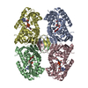
 PDBj
PDBj Assembly
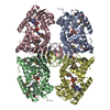

 Mass: 663.425 Da / Num. of mol.: 4 / Source method: obtained synthetically / Formula: C21H27N7O14P2 / Comment: NAD*YM
 Mass: 35.453 Da / Num. of mol.: 1 / Source method: obtained synthetically / Formula: Cl
 Mass: 62.068 Da / Num. of mol.: 3 / Source method: obtained synthetically / Formula: C2H6O2
 Mass: 18.015 Da / Num. of mol.: 343 / Source method: isolated from a natural source / Formula: H2O
 Sample preparation
 / Beamline: BL11-1 / Wavelength: 0.91837, 0.97941, 0.97883
 Processing