| 登録情報 | データベース: PDB / ID: 1xdz
|
|---|
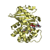
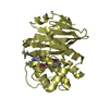
| タイトル | Crystal Structure of Gram_Positive Bacillus subtilis Glucose inhibited Division protein B (gidB), Structural genomics, MCSG |
|---|
 要素 要素 | Methyltransferase gidB |
|---|
 キーワード キーワード | TRANSFERASE / MCSG / Protein Structure Initiative / Structural Genomics / methyltransferase fold / GidB / PSI / Midwest Center for Structural Genomics |
|---|
| 機能・相同性 |  機能・相同性情報 機能・相同性情報
rRNA (guanine-N7-)-methyltransferase activity / 転移酵素; 一炭素原子の基を移すもの; メチル基を移すもの / cytosol類似検索 - 分子機能 rRNA small subunit methyltransferase G / rRNA small subunit methyltransferase G / Vaccinia Virus protein VP39 / S-adenosyl-L-methionine-dependent methyltransferase superfamily / Rossmann fold / 3-Layer(aba) Sandwich / Alpha Beta類似検索 - ドメイン・相同性 Ribosomal RNA small subunit methyltransferase G類似検索 - 構成要素 |
|---|
| 生物種 |   Bacillus subtilis (枯草菌) Bacillus subtilis (枯草菌) |
|---|
| 手法 |  X線回折 / シンクロトロン / 単波長異常分散 / 解像度: 1.6 Å X線回折 / シンクロトロン / 単波長異常分散 / 解像度: 1.6 Å |
|---|
 データ登録者 データ登録者 | Zhang, R. / Wu, R. / Collart, F. / Joachimiak, A. / Midwest Center for Structural Genomics (MCSG) |
|---|
 引用 引用 | ジャーナル: To be Published
タイトル: The 1.6A crystal ctructure of Gram-positive Bacillus subtilis glucose inhibited division protein B (gidB)
著者: Zhang, R. / Wu, R. / Collart, F. / Joachimiak, A. |
|---|
| 履歴 | | 登録 | 2004年9月8日 | 登録サイト: RCSB / 処理サイト: RCSB |
|---|
| 改定 1.0 | 2004年10月26日 | Provider: repository / タイプ: Initial release |
|---|
| 改定 1.1 | 2008年4月30日 | Group: Version format compliance |
|---|
| 改定 1.2 | 2011年7月13日 | Group: Version format compliance |
|---|
| 改定 1.3 | 2024年2月14日 | Group: Data collection / Database references
カテゴリ: chem_comp_atom / chem_comp_bond ...chem_comp_atom / chem_comp_bond / database_2 / struct_ref_seq_dif
Item: _database_2.pdbx_DOI / _database_2.pdbx_database_accession / _struct_ref_seq_dif.details |
|---|
|
|---|
 ムービー
ムービー コントローラー


 データを開く
 基本情報
基本情報 構造の表示
構造の表示 ダウンロードとリンク
 その他のダウンロード
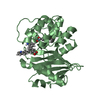


 PDBj
PDBj 集合体
集合体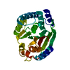
 分子量: 18.015 Da / 分子数: 211 / 由来タイプ: 天然 / 式: H2O
 試料調製
試料調製 / ビームライン: 19-ID / 波長: 0.97835 Å
 解析
解析