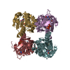
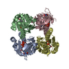
| Deposited unit | A: Thymidine kinase, cytosolic
B: Thymidine kinase, cytosolic
C: Thymidine kinase, cytosolic
D: Thymidine kinase, cytosolic
E: Thymidine kinase, cytosolic
F: Thymidine kinase, cytosolic
G: Thymidine kinase, cytosolic
H: Thymidine kinase, cytosolic
hetero molecules
| Theoretical mass | Number of molelcules |
|---|
| Total (without water) | 175,191 | 32 |
|---|
| Polymers | 170,616 | 8 |
|---|
| Non-polymers | 4,575 | 24 |
|---|
| Water | 3,837 | 213 |
|---|
|
|---|
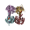
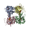
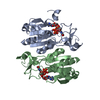
| 1 | A: Thymidine kinase, cytosolic
B: Thymidine kinase, cytosolic
C: Thymidine kinase, cytosolic
D: Thymidine kinase, cytosolic
hetero molecules
| Theoretical mass | Number of molelcules |
|---|
| Total (without water) | 87,596 | 16 |
|---|
| Polymers | 85,308 | 4 |
|---|
| Non-polymers | 2,288 | 12 |
|---|
| Water | 72 | 4 |
|---|
| Type | Name | Symmetry operation | Number |
|---|
| identity operation | 1_555 | x,y,z | 1 |
| Buried area | 12360 Å2 |
|---|
| ΔGint | -93 kcal/mol |
|---|
| Surface area | 25830 Å2 |
|---|
| Method | PISA |
|---|
|
|---|
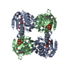
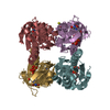
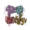
| 2 | E: Thymidine kinase, cytosolic
F: Thymidine kinase, cytosolic
G: Thymidine kinase, cytosolic
H: Thymidine kinase, cytosolic
hetero molecules
| Theoretical mass | Number of molelcules |
|---|
| Total (without water) | 87,596 | 16 |
|---|
| Polymers | 85,308 | 4 |
|---|
| Non-polymers | 2,288 | 12 |
|---|
| Water | 72 | 4 |
|---|
| Type | Name | Symmetry operation | Number |
|---|
| identity operation | 1_555 | x,y,z | 1 |
| Buried area | 12430 Å2 |
|---|
| ΔGint | -98 kcal/mol |
|---|
| Surface area | 26040 Å2 |
|---|
| Method | PISA |
|---|
|
|---|
| Unit cell | | Length a, b, c (Å) | 157.672, 123.177, 116.000 |
|---|
| Angle α, β, γ (deg.) | 90.00, 130.20, 90.00 |
|---|
| Int Tables number | 5 |
|---|
| Space group name H-M | C121 |
|---|
|
|---|
| Noncrystallographic symmetry (NCS) | NCS domain: | ID | Ens-ID | Details (eV) |
|---|
| 1 | 1 | A| 2 | 1 | B| 3 | 1 | C| 4 | 1 | D| 5 | 1 | E| 6 | 1 | F| 7 | 1 | G| 8 | 1 | H| 9 | 1 | A| 10 | 1 | B| 11 | 1 | C| 12 | 1 | D| 13 | 1 | E| 14 | 1 | F| 15 | 1 | G| 16 | 1 | H | | | | | | | | | | | | | | | |
NCS domain segments: Ens-ID: 1 / Refine code: 2 | Dom-ID | Component-ID | Beg auth comp-ID | Beg label comp-ID | End auth comp-ID | End label comp-ID | Auth asym-ID | Label asym-ID | Auth seq-ID | Label seq-ID |
|---|
| 1 | 1 | ARGARGASPASPAA| 18 - 58 | 18 - 58 | | 2 | 1 | ARGARGASPASPBB| 18 - 58 | 18 - 58 | | 3 | 1 | ARGARGASPASPCC| 18 - 58 | 18 - 58 | | 4 | 1 | ARGARGASPASPDD| 18 - 58 | 18 - 58 | | 5 | 1 | ARGARGASPASPEE| 18 - 58 | 18 - 58 | | 6 | 1 | ARGARGASPASPFF| 18 - 58 | 18 - 58 | | 7 | 1 | ARGARGASPASPGG| 18 - 58 | 18 - 58 | | 8 | 1 | ARGARGASPASPHH| 18 - 58 | 18 - 58 | | 9 | 2 | ALAALALYSLYSAA| 75 - 191 | 75 - 191 | | 10 | 2 | ALAALALYSLYSBB| 75 - 191 | 75 - 191 | | 11 | 2 | ALAALALYSLYSCC| 75 - 191 | 75 - 191 | | 12 | 2 | ALAALALYSLYSDD| 75 - 191 | 75 - 191 | | 13 | 2 | ALAALALYSLYSE| E | | | | | | | | | | | | | | | | | | | | | | | | | | | | | | | | | | | | | | | | | | | | | | | | | | | | | | | | | | | | | | | | | | | | | | | | | | | | | |
|
|---|
 Movie
Movie Controller


 Open data
 Basic information
 Components
 Keywords
 Function and homology information
 Homo sapiens (human)
 X-RAY DIFFRACTION /
  Authors
 Citation
 Structure visualization
 Downloads & links
 Other downloads
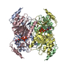
 PDBj
PDBj


 Assembly