 Movie
Movie Controller

[English] 日本語
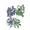
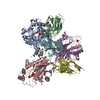
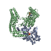
 Yorodumi
- PDB-1x6v: The crystal structure of human 3'-phosphoadenosine-5'-phosphosulf... -
+ Open data

- Basic information
| Entry | Database: PDB / ID: 1x6v | ||||||
|---|---|---|---|---|---|---|---|
| Title | The crystal structure of human 3'-phosphoadenosine-5'-phosphosulfate synthetase 1 | ||||||
 Components Components | Bifunctional 3'-phosphoadenosine 5'-phosphosulfate synthetase 1 | ||||||
 Keywords Keywords | TRANSFERASE / ATP sulfurylase / APS kinase / PAPS / phosphoadenosine phosphosulfate | ||||||
| Function / homology |  Function and homology information Function and homology information3'-phosphoadenosine 5'-phosphosulfate biosynthetic process / Transport and metabolism of PAPS / sulfate adenylyltransferase / adenylyl-sulfate kinase / adenylylsulfate kinase activity / sulfate adenylyltransferase (ATP) activity / Metabolism of ingested H2SeO4 and H2SeO3 into H2Se / sulfate assimilation / nucleotidyltransferase activity / skeletal system development ...3'-phosphoadenosine 5'-phosphosulfate biosynthetic process / Transport and metabolism of PAPS / sulfate adenylyltransferase / adenylyl-sulfate kinase / adenylylsulfate kinase activity / sulfate adenylyltransferase (ATP) activity / Metabolism of ingested H2SeO4 and H2SeO3 into H2Se / sulfate assimilation / nucleotidyltransferase activity / skeletal system development / Signaling by BRAF and RAF1 fusions / protein homodimerization activity / ATP binding / nucleus / cytosol Similarity search - Function | ||||||
| Biological species |  Homo sapiens (human) Homo sapiens (human) | ||||||
| Method |  X-RAY DIFFRACTION / SYNCHROTRON / MOLECULAR REPLACEMENT WITH INITIAL MERCURY PHASES / Resolution: 1.75 Å X-RAY DIFFRACTION / SYNCHROTRON / MOLECULAR REPLACEMENT WITH INITIAL MERCURY PHASES / Resolution: 1.75 Å | ||||||
 Authors Authors | Harjes, S. / Bayer, P. / Scheidig, A.J. | ||||||
 Citation Citation | Journal: J.Mol.Biol. / Year: 2005 Title: The Crystal Structure of Human PAPS Synthetase 1 Reveals Asymmetry in Substrate Binding Authors: Harjes, S. / Bayer, P. / Scheidig, A.J. | ||||||
| History |
|
- Structure visualization
| Structure viewer | Molecule: MolmilJmol/JSmol |
|---|
- Downloads & links
-Download
| PDBx/mmCIF format | 1x6v.cif.gz | 265.9 KB | Display | PDBx/mmCIF format |
|---|---|---|---|---|
| PDB format | pdb1x6v.ent.gz | 209.5 KB | Display | PDB format |
| PDBx/mmJSON format | 1x6v.json.gz | Tree view | PDBx/mmJSON format | |
| Others |  Other downloads Other downloads |
-Validation report
| Arichive directory | https://data.pdbj.org/pub/pdb/validation_reports/x6/1x6vftp://data.pdbj.org/pub/pdb/validation_reports/x6/1x6v | HTTPS FTP |
|---|
-Related structure data
| Related structure data |  1xjqC  1xnjC  1d6jS  1g8fS S: Starting model for refinement C: citing same article ( |
|---|---|
| Similar structure data |


-Links
 PDBj
PDBj
- Assembly
| Deposited unit | 
| ||||||||
|---|---|---|---|---|---|---|---|---|---|
| 1 |
| ||||||||
| Unit cell |
|
-Components
| #1: Protein | Mass: 71698.945 Da / Num. of mol.: 2 Source method: isolated from a genetically manipulated source Details: [Includes: Sulfate adenylyltransferase (synonym: Sulfate adenylate transferase, SAT, ATP-sulfurylase); Adenylyl-sulfate kinase (synonym: Adenylylsulfate 3'-phosphotransferase, APS kinase, ...Details: [Includes: Sulfate adenylyltransferase (synonym: Sulfate adenylate transferase, SAT, ATP-sulfurylase); Adenylyl-sulfate kinase (synonym: Adenylylsulfate 3'-phosphotransferase, APS kinase, Adenosine-5'-phosphosulfate 3'-phosphotransferase, 3'- phosphoadenosine-5'-phosphosulfate synthetase)] Source: (gene. exp.) Homo sapiens (human) / Gene: PAPSS1, PAPSS, ATPSK1 / Plasmid: pET27bmod / Species (production host): Escherichia coli / Production host:  References: UniProt: O43252, sulfate adenylyltransferase, adenylyl-sulfate kinase #2: Chemical |   Mass: 35.453 Da / Num. of mol.: 2 / Source method: obtained synthetically / Formula: Cl Mass: 35.453 Da / Num. of mol.: 2 / Source method: obtained synthetically / Formula: Cl#3: Chemical | ChemComp-ADP / |   Mass: 427.201 Da / Num. of mol.: 1 / Source method: obtained synthetically / Formula: C10H15N5O10P2 / Comment: ADP, energy-carrying molecule*YM Mass: 427.201 Da / Num. of mol.: 1 / Source method: obtained synthetically / Formula: C10H15N5O10P2 / Comment: ADP, energy-carrying molecule*YM#4: Water | ChemComp-HOH / |  Mass: 18.015 Da / Num. of mol.: 1091 / Source method: isolated from a natural source / Formula: H2O Mass: 18.015 Da / Num. of mol.: 1091 / Source method: isolated from a natural source / Formula: H2O |
|---|
-Experimental details
-Experiment
| Experiment | Method: X-RAY DIFFRACTION / Number of used crystals: 1 |
|---|
- Sample preparation
| Crystal | Density Matthews: 2.9 Å3/Da / Density % sol: 57 % |
|---|---|
| Crystal grow | Temperature: 285 K / pH: 5.6 Details: 91 mM sodium Citrate, 270 mM ammonium acetate, 12% PEG 4000, pH 5.6, VAPOR DIFFUSION, HANGING DROP, temperature 285K |
-Data collection
| Diffraction | Mean temperature: 100 K |
|---|---|
| Diffraction source | Source: SYNCHROTRON / Site: ESRF  / Beamline: ID14-1 / Wavelength: 0.934 / Beamline: ID14-1 / Wavelength: 0.934 |
| Detector | Type: ADSC QUANTUM 4 / Detector: CCD / Date: Mar 1, 2003 |
| Radiation | Protocol: SINGLE WAVELENGTH / Monochromatic (M) / Laue (L): M / Scattering type: x-ray |
| Radiation wavelength | Wavelength: 0.934 Å / Relative weight: 1 |
| Reflection | Resolution: 1.75→30 Å / Num. obs: 159267 / Observed criterion σ(I): 1 / Redundancy: 3.6 % / Rsym value: 0.069 / Net I/σ(I): 11 |
| Reflection shell | Resolution: 1.75→1.85 Å / Redundancy: 3.2 % / Mean I/σ(I) obs: 2.1 / Rsym value: 0.58 / % possible all: 88 |
- Processing
| Software |
| ||||||||||||||||||||||||||||||||||||||||||||||||||||||||||||||||||||||||||||||||||||||||||||||||||||||||||||||||||||||||||||||||||||||||||||||||||||||||||||||||||||||||||
|---|---|---|---|---|---|---|---|---|---|---|---|---|---|---|---|---|---|---|---|---|---|---|---|---|---|---|---|---|---|---|---|---|---|---|---|---|---|---|---|---|---|---|---|---|---|---|---|---|---|---|---|---|---|---|---|---|---|---|---|---|---|---|---|---|---|---|---|---|---|---|---|---|---|---|---|---|---|---|---|---|---|---|---|---|---|---|---|---|---|---|---|---|---|---|---|---|---|---|---|---|---|---|---|---|---|---|---|---|---|---|---|---|---|---|---|---|---|---|---|---|---|---|---|---|---|---|---|---|---|---|---|---|---|---|---|---|---|---|---|---|---|---|---|---|---|---|---|---|---|---|---|---|---|---|---|---|---|---|---|---|---|---|---|---|---|---|---|---|---|---|---|
| Refinement | Method to determine structure: MOLECULAR REPLACEMENT WITH INITIAL MERCURY PHASES Starting model: PDB ENTRIES 1G8F, 1D6J Resolution: 1.75→129.1 Å / Cor.coef. Fo:Fc: 0.97 / Cor.coef. Fo:Fc free: 0.958 / SU B: 2.311 / SU ML: 0.07 / TLS residual ADP flag: LIKELY RESIDUAL / Cross valid method: THROUGHOUT / ESU R: 0.089 / ESU R Free: 0.091 / Stereochemistry target values: MAXIMUM LIKELIHOOD / Details: HYDROGENS HAVE BEEN ADDED IN THE RIDING POSITIONS
| ||||||||||||||||||||||||||||||||||||||||||||||||||||||||||||||||||||||||||||||||||||||||||||||||||||||||||||||||||||||||||||||||||||||||||||||||||||||||||||||||||||||||||
| Solvent computation | Ion probe radii: 0.8 Å / Shrinkage radii: 0.8 Å / VDW probe radii: 1.4 Å / Solvent model: BABINET MODEL WITH MASK | ||||||||||||||||||||||||||||||||||||||||||||||||||||||||||||||||||||||||||||||||||||||||||||||||||||||||||||||||||||||||||||||||||||||||||||||||||||||||||||||||||||||||||
| Displacement parameters | Biso mean: 24.273 Å2
| ||||||||||||||||||||||||||||||||||||||||||||||||||||||||||||||||||||||||||||||||||||||||||||||||||||||||||||||||||||||||||||||||||||||||||||||||||||||||||||||||||||||||||
| Refinement step | Cycle: LAST / Resolution: 1.75→129.1 Å
| ||||||||||||||||||||||||||||||||||||||||||||||||||||||||||||||||||||||||||||||||||||||||||||||||||||||||||||||||||||||||||||||||||||||||||||||||||||||||||||||||||||||||||
| Refine LS restraints |
| ||||||||||||||||||||||||||||||||||||||||||||||||||||||||||||||||||||||||||||||||||||||||||||||||||||||||||||||||||||||||||||||||||||||||||||||||||||||||||||||||||||||||||
| LS refinement shell | Resolution: 1.75→1.795 Å / Total num. of bins used: 20 /
| ||||||||||||||||||||||||||||||||||||||||||||||||||||||||||||||||||||||||||||||||||||||||||||||||||||||||||||||||||||||||||||||||||||||||||||||||||||||||||||||||||||||||||
| Refinement TLS params. | Method: refined / Refine-ID: X-RAY DIFFRACTION
| ||||||||||||||||||||||||||||||||||||||||||||||||||||||||||||||||||||||||||||||||||||||||||||||||||||||||||||||||||||||||||||||||||||||||||||||||||||||||||||||||||||||||||
| Refinement TLS group |
|
