- PDB-1x3s: Crystal structure of human Rab18 in complex with Gppnhp -
+
Open data
ID or keywords:
Loading...
-
Basic information
Entry
Database: PDB / ID: 1x3s
Title
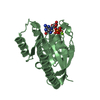
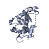
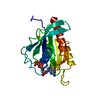
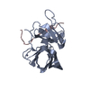
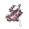
Crystal structure of human Rab18 in complex with Gppnhp
Components
Ras-related protein Rab-18
Keywords
ENDOCYTOSIS/EXOCYTOSIS / GTPase / Rab / GNP / Structural Genomics / NPPSFA / National Project on Protein Structural and Functional Analyses / RIKEN Structural Genomics/Proteomics Initiative / RSGI / ENDOCYTOSIS-EXOCYTOSIS COMPLEX
Function / homology
Function and homology information
endoplasmic reticulum tubular network organization / endoplasmic reticulum tubular network / eye development / RAB geranylgeranylation / cis-Golgi network membrane / lipid droplet organization / RAB GEFs exchange GTP for GDP on RABs / COPI-independent Golgi-to-ER retrograde traffic / small GTPase-mediated signal transduction / endomembrane system ...endoplasmic reticulum tubular network organization / endoplasmic reticulum tubular network / eye development / RAB geranylgeranylation / cis-Golgi network membrane / lipid droplet organization / RAB GEFs exchange GTP for GDP on RABs / COPI-independent Golgi-to-ER retrograde traffic / small GTPase-mediated signal transduction / endomembrane system / lipid droplet / secretory granule membrane / small monomeric GTPase / brain development / intracellular protein transport / GDP binding / G protein activity / apical plasma membrane / GTPase activity / Neutrophil degranulation / endoplasmic reticulum membrane / GTP binding / Golgi apparatus / plasma membrane / cytosol Similarity search - Function
: / Sigma-54 interaction domain, ATP-binding site 1 / ARF-like small GTPases; ARF, ADP-ribosylation factor / Small GTPase Rab domain profile. / Ran (Ras-related nuclear proteins) /TC4 subfamily of small GTPases / Rho (Ras homology) subfamily of Ras-like small GTPases / Ras subfamily of RAS small GTPases / Small GTPase / Ras family / Rab subfamily of small GTPases ...: / Sigma-54 interaction domain, ATP-binding site 1 / ARF-like small GTPases; ARF, ADP-ribosylation factor / Small GTPase Rab domain profile. / Ran (Ras-related nuclear proteins) /TC4 subfamily of small GTPases / Rho (Ras homology) subfamily of Ras-like small GTPases / Ras subfamily of RAS small GTPases / Small GTPase / Ras family / Rab subfamily of small GTPases / Small GTP-binding protein domain / P-loop containing nucleotide triphosphate hydrolases / Rossmann fold / P-loop containing nucleoside triphosphate hydrolase / 3-Layer(aba) Sandwich / Alpha Beta Similarity search - Domain/homology
In the structure databanks used in Yorodumi, some data are registered as the other names, "COVID-19 virus" and "2019-nCoV". Here are the details of the virus and the list of structure data.
Jan 31, 2019. EMDB accession codes are about to change! (news from PDBe EMDB page)
EMDB accession codes are about to change! (news from PDBe EMDB page)
The allocation of 4 digits for EMDB accession codes will soon come to an end. Whilst these codes will remain in use, new EMDB accession codes will include an additional digit and will expand incrementally as the available range of codes is exhausted. The current 4-digit format prefixed with “EMD-” (i.e. EMD-XXXX) will advance to a 5-digit format (i.e. EMD-XXXXX), and so on. It is currently estimated that the 4-digit codes will be depleted around Spring 2019, at which point the 5-digit format will come into force.
The EM Navigator/Yorodumi systems omit the EMD- prefix.
Related info.:Q: What is EMD? / ID/Accession-code notation in Yorodumi/EM Navigator
Yorodumi is a browser for structure data from EMDB, PDB, SASBDB, etc.
This page is also the successor to EM Navigator detail page, and also detail information page/front-end page for Omokage search.
The word "yorodu" (or yorozu) is an old Japanese word meaning "ten thousand". "mi" (miru) is to see.
Related info.:EMDB / PDB / SASBDB / Comparison of 3 databanks / Yorodumi Search / Aug 31, 2016. New EM Navigator & Yorodumi / Yorodumi Papers / Jmol/JSmol / Function and homology information / Changes in new EM Navigator and Yorodumi
 Movie
Movie Controller


 Open data
 Basic information
 Components
 Keywords
 Function and homology information
 Homo sapiens (human)
 X-RAY DIFFRACTION /
  Authors
 Citation
 Structure visualization
 Downloads & links
 Other downloads

 PDBj
PDBj


 Assembly

 Mass: 24.305 Da / Num. of mol.: 1 / Source method: obtained synthetically / Formula: Mg
 Mass: 522.196 Da / Num. of mol.: 1 / Source method: obtained synthetically / Formula: C10H17N6O13P3
 Mass: 18.015 Da / Num. of mol.: 230 / Source method: isolated from a natural source / Formula: H2O
 Sample preparation
 / Beamline: 22-ID / Wavelength: 0.97931, 0.97917, 0.96860
 Processing