 Movie
Movie Controller

[English] 日本語
 Yorodumi
- PDB-1x1l: Interaction of ERA,a GTPase protein, with the 3'minor domain of t... -
+ Open data

- Basic information
| Entry | Database: PDB / ID: 1x1l | ||||||
|---|---|---|---|---|---|---|---|
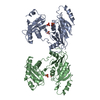
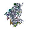
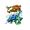
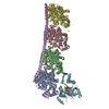
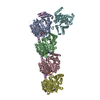
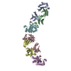
| Title | Interaction of ERA,a GTPase protein, with the 3'minor domain of the 16S rRNA within the THERMUS THERMOPHILUS 30S subunit. | ||||||
 Components Components |
| ||||||
 Keywords Keywords | STRUCTURAL PROTEIN/RNA / Interaction of Era protein with the 3'minor domain of 16S rRNA / STRUCTURAL PROTEIN-RNA COMPLEX | ||||||
| Function / homology |  Function and homology information Function and homology informationguanosine tetraphosphate binding / ribosomal small subunit binding / ribosomal small subunit assembly / ribosomal small subunit biogenesis / small ribosomal subunit rRNA binding / protein phosphorylation / rRNA binding / GTPase activity / GTP binding / RNA binding ...guanosine tetraphosphate binding / ribosomal small subunit binding / ribosomal small subunit assembly / ribosomal small subunit biogenesis / small ribosomal subunit rRNA binding / protein phosphorylation / rRNA binding / GTPase activity / GTP binding / RNA binding / plasma membrane / cytoplasm / cytosol Similarity search - Function | ||||||
| Biological species |   Thermus thermophilus (bacteria) Thermus thermophilus (bacteria) | ||||||
| Method | ELECTRON MICROSCOPY / single particle reconstruction / cryo EM / Resolution: 13.5 Å | ||||||
 Authors Authors | Sharma, M.R. / Barat, C. / Agrawal, R.K. | ||||||
 Citation Citation | Journal: Mol Cell / Year: 2005 Title: Interaction of Era with the 30S ribosomal subunit implications for 30S subunit assembly. Authors: Manjuli R Sharma / Chandana Barat / Daniel N Wilson / Timothy M Booth / Masahito Kawazoe / Chie Hori-Takemoto / Mikako Shirouzu / Shigeyuki Yokoyama / Paola Fucini / Rajendra K Agrawal /  Abstract: Era (E. coliRas-like protein) is a highly conserved and essential GTPase in bacteria. It binds to the 16S ribosomal RNA (rRNA) of the small (30S) ribosomal subunit, and its depletion leads to ...Era (E. coliRas-like protein) is a highly conserved and essential GTPase in bacteria. It binds to the 16S ribosomal RNA (rRNA) of the small (30S) ribosomal subunit, and its depletion leads to accumulation of an unprocessed precursor of the 16S rRNA. We have obtained a three-dimensional cryo-electron microscopic map of the Thermus thermophilus 30S-Era complex. Era binds in the cleft between the head and platform of the 30S subunit and locks the subunit in a conformation that is not favorable for association with the large (50S) ribosomal subunit. The RNA binding KH motif present within the C-terminal domain of Era interacts with the conserved nucleotides in the 3' region of the 16S rRNA. Furthermore, Era makes contact with several assembly elements of the 30S subunit. These observations suggest a direct involvement of Era in the assembly and maturation of the 30S subunit. #1: Journal: Nature / Year: 2000 Title: Molecular biology. Small subunit, big science. #2: Journal: Proc Natl Acad Sci U S A / Year: 1999Title: Crystal structure of ERA: a GTPase-dependent cell cycle regulator containing an RNA binding motif. Authors: X Chen / D L Court / X Ji / Abstract: ERA forms a unique family of GTPase. It is widely conserved and essential in bacteria. ERA functions in cell cycle control by coupling cell division with growth rate. ERA homologues also are found in ...ERA forms a unique family of GTPase. It is widely conserved and essential in bacteria. ERA functions in cell cycle control by coupling cell division with growth rate. ERA homologues also are found in eukaryotes. Here we report the crystal structure of ERA from Escherichia coli. The structure has been determined at 2.4-A resolution. It reveals a two-domain arrangement of the molecule: an N-terminal domain that resembles p21 Ras and a C-terminal domain that is unique. Structure-based topological search of the C domain fails to reveal any meaningful match, although sequence analysis suggests that it contains a KH domain. KH domains are RNA binding motifs that usually occur in tandem repeats and exhibit low sequence similarity except for the well-conserved segment VIGxxGxxIK. We have identified a betaalphaalphabeta fold that contains the VIGxxGxxIK sequence and is shared by the C domain of ERA and the KH domain. We propose that this betaalphaalphabeta fold is the RNA binding motif, the minimum structural requirement for RNA binding. ERA dimerizes in crystal. The dimer formation involves a significantly distorted switch II region, which may shed light on how ERA protein regulates downstream events. #3: Journal: To be PublishedTitle: Crystal structure of Era from Thermus thermophilus Authors: Kawazoe, M. / Takemoto, C. / Kaminishi, T. / Sekine, S. / Shirouzu, M. / Fucini, P. / Agrawal, R.K. / Yokoyama, S. | ||||||
| History |
|
- Structure visualization
| Movie |
Movie viewer |
|---|---|
| Structure viewer | Molecule: MolmilJmol/JSmol |
- Downloads & links
-Download
| PDBx/mmCIF format | 1x1l.cif.gz | 29.2 KB | Display | PDBx/mmCIF format |
|---|---|---|---|---|
| PDB format | pdb1x1l.ent.gz | 13.6 KB | Display | PDB format |
| PDBx/mmJSON format | 1x1l.json.gz | Tree view | PDBx/mmJSON format | |
| Others |  Other downloads Other downloads |
-Validation report
| Arichive directory | https://data.pdbj.org/pub/pdb/validation_reports/x1/1x1lftp://data.pdbj.org/pub/pdb/validation_reports/x1/1x1l | HTTPS FTP |
|---|
-Related structure data


-Links
 PDBj
PDBj


- Assembly
| Deposited unit |
|
|---|---|
| 1 |
|
-Components
| #1: RNA chain | Mass: 42303.242 Da / Num. of mol.: 1 / Source method: isolated from a natural source / Source: (natural) Thermus thermophilus (bacteria) |
|---|---|
| #2: Protein | Mass: 33858.078 Da / Num. of mol.: 1 / Source method: isolated from a natural source / Source: (natural) |
-Experimental details
-Experiment
| Experiment | Method: ELECTRON MICROSCOPY |
|---|---|
| EM experiment | Aggregation state: PARTICLE / 3D reconstruction method: single particle reconstruction |
- Sample preparation
| Component | Name: THERMUS THERMOPHILUS 30S ribosomal subunit complexed with Era Type: RIBOSOME / Details: Era was bound to a S1-depleted 30S subunit |
|---|---|
| Buffer solution | Name: Hepes-KOH / pH: 7.5 / Details: Hepes-KOH |
| Specimen | Conc.: 0.032 mg/ml / Embedding applied: NO / Shadowing applied: NO / Staining applied: NO / Vitrification applied: YES |
| Specimen support | Details: Quantifoil holley-carbon film GRIDS |
| Vitrification | Details: Rapid-freezing in liquid ethane |
- Electron microscopy imaging
| Experimental equipment |  Model: Tecnai F20 / Image courtesy: FEI Company |
|---|---|
| Microscopy | Model: FEI TECNAI F20 / Date: Mar 25, 2003 |
| Electron gun | Electron source:  FIELD EMISSION GUN / Accelerating voltage: 200 kV / Illumination mode: FLOOD BEAM FIELD EMISSION GUN / Accelerating voltage: 200 kV / Illumination mode: FLOOD BEAM |
| Electron lens | Mode: BRIGHT FIELD / Nominal magnification: 50000 X / Calibrated magnification: 49696 X / Nominal defocus max: 3940 nm / Nominal defocus min: 1180 nm / Cs: 2 mm |
| Specimen holder | Temperature: 93 K / Tilt angle max: 0 ° / Tilt angle min: 0 ° |
| Image recording | Electron dose: 20 e/Å2 / Film or detector model: KODAK SO-163 FILM |
| Radiation | Protocol: SINGLE WAVELENGTH / Monochromatic (M) / Laue (L): M / Scattering type: x-ray |
| Radiation wavelength | Relative weight: 1 |
- Processing
| EM software |
| |||||||||||||||||||||
|---|---|---|---|---|---|---|---|---|---|---|---|---|---|---|---|---|---|---|---|---|---|---|
| CTF correction | Details: CTF correction of 3D-maps by wiener filtration | |||||||||||||||||||||
| Symmetry | Point symmetry: C1 (asymmetric) | |||||||||||||||||||||
| 3D reconstruction | Method: reference based alignment / Resolution: 13.5 Å / Actual pixel size: 2.82 Å / Magnification calibration: TMV Details: projection matching using spider package. The coordinates for only the alpha carbons in protein and phosphoruses in rRNA are present in the structure. The number of missing atoms was so much ...Details: projection matching using spider package. The coordinates for only the alpha carbons in protein and phosphoruses in rRNA are present in the structure. The number of missing atoms was so much that remark 470 for the missing atoms list were removed. Symmetry type: POINT | |||||||||||||||||||||
| Atomic model building | Protocol: RIGID BODY FIT / Space: REAL Target criteria: X-ray coordinates of the 30S ribosomal subunit and ERA were fitted into the 13.5 angstroms resolution CRYO-EM map of the T. Thermophilus 30S subunit-ERA complex. the atomic structure ...Target criteria: X-ray coordinates of the 30S ribosomal subunit and ERA were fitted into the 13.5 angstroms resolution CRYO-EM map of the T. Thermophilus 30S subunit-ERA complex. the atomic structure of ERA was fitted as 3 rigid bodies, N-terminal domain, C-terminal domain and C-terminal helix within the C-terminal domain. The resultant ERA structure was then energy minimized. The X-ray coordinates of T. Thermophilus 30S subunit was fitted as 4 rigid bodies, head, body, platform and 16S rRNA 3' minor domains. Only the coordinates of the 16S rRNA 3' minor domain and ERA are included HERE. The KH domain of ERA makes direct contact with the 3' terminus of the 16S rRNA. Details: METHOD--cross-correlation based manual fitting in O REFINEMENT PROTOCOL--MULTIPLE RIGID BODY | |||||||||||||||||||||
| Atomic model building |
| |||||||||||||||||||||
| Refinement step | Cycle: LAST
|

