| 登録情報 | データベース: PDB / ID: 1x1c
|
|---|
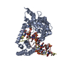
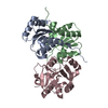
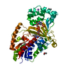
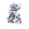
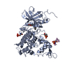
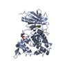
| タイトル | Crystal structure of BchU complexed with S-adenosyl-L-homocysteine and Zn2+ |
|---|
 要素 要素 | CrtF-related protein |
|---|
 キーワード キーワード | TRANSFERASE / METHYLTRANSFERASE / BACTERIOCHLLOCHLOROPHYLL / BchU / SAM / SAH / S-ADENOSYLMETHYONINE / S-ADENOSYLHOMOCYSTEINE / ADO-MET / ADO-HCY |
|---|
| 機能・相同性 |  機能・相同性情報 機能・相同性情報
bacteriochlorophyllide d C-20 methyltransferase / light-dependent bacteriochlorophyll biosynthetic process / light-independent bacteriochlorophyll biosynthetic process / O-methyltransferase activity / methyltransferase activity / methylation / protein dimerization activity / metal ion binding類似検索 - 分子機能 C-20 methyltransferase CrtF-related / Plant methyltransferase dimerisation / O-methyltransferase dimerisation domain / O-methyltransferase domain / O-methyltransferase COMT-type / O-methyltransferase domain / SAM-dependent O-methyltransferase class II-type profile. / Vaccinia Virus protein VP39 / Winged helix-like DNA-binding domain superfamily/Winged helix DNA-binding domain / Arc Repressor Mutant, subunit A ...C-20 methyltransferase CrtF-related / Plant methyltransferase dimerisation / O-methyltransferase dimerisation domain / O-methyltransferase domain / O-methyltransferase COMT-type / O-methyltransferase domain / SAM-dependent O-methyltransferase class II-type profile. / Vaccinia Virus protein VP39 / Winged helix-like DNA-binding domain superfamily/Winged helix DNA-binding domain / Arc Repressor Mutant, subunit A / Winged helix DNA-binding domain superfamily / Winged helix-like DNA-binding domain superfamily / S-adenosyl-L-methionine-dependent methyltransferase superfamily / Rossmann fold / Orthogonal Bundle / 3-Layer(aba) Sandwich / Mainly Alpha / Alpha Beta類似検索 - ドメイン・相同性 S-ADENOSYL-L-HOMOCYSTEINE / Bacteriochlorophyllide d C-20 methyltransferase類似検索 - 構成要素 |
|---|
| 生物種 |  Chlorobium tepidum (バクテリア) Chlorobium tepidum (バクテリア) |
|---|
| 手法 |  X線回折 / シンクロトロン / 分子置換 / 解像度: 2.85 Å X線回折 / シンクロトロン / 分子置換 / 解像度: 2.85 Å |
|---|
 データ登録者 データ登録者 | Yamaguchi, H. / Wada, K. / Fukuyama, K. |
|---|
 引用 引用 | ジャーナル: J.Mol.Biol. / 年: 2006
タイトル: Crystal Structures of BchU, a Methyltransferase Involved in Bacteriochlorophyll c Biosynthesis, and its Complex with S-adenosylhomocysteine: Implications for Reaction Mechanism.
著者: Wada, K. / Yamaguchi, H. / Harada, J. / Niimi, K. / Osumi, S. / Saga, Y. / Oh-Oka, H. / Tamiaki, H. / Fukuyama, K. |
|---|
| 履歴 | | 登録 | 2005年4月3日 | 登録サイト: PDBJ / 処理サイト: PDBJ |
|---|
| 改定 1.0 | 2006年7月18日 | Provider: repository / タイプ: Initial release |
|---|
| 改定 1.1 | 2008年4月30日 | Group: Version format compliance |
|---|
| 改定 1.2 | 2011年7月13日 | Group: Derived calculations / Source and taxonomy / Version format compliance |
|---|
| 改定 1.3 | 2023年10月25日 | Group: Data collection / Database references ...Data collection / Database references / Derived calculations / Refinement description
カテゴリ: chem_comp_atom / chem_comp_bond ...chem_comp_atom / chem_comp_bond / database_2 / pdbx_initial_refinement_model / pdbx_struct_conn_angle / struct_conn / struct_ref_seq_dif / struct_site
Item: _database_2.pdbx_DOI / _database_2.pdbx_database_accession ..._database_2.pdbx_DOI / _database_2.pdbx_database_accession / _pdbx_struct_conn_angle.ptnr1_auth_comp_id / _pdbx_struct_conn_angle.ptnr1_auth_seq_id / _pdbx_struct_conn_angle.ptnr1_label_atom_id / _pdbx_struct_conn_angle.ptnr1_label_comp_id / _pdbx_struct_conn_angle.ptnr1_label_seq_id / _pdbx_struct_conn_angle.ptnr1_symmetry / _pdbx_struct_conn_angle.ptnr2_auth_seq_id / _pdbx_struct_conn_angle.ptnr2_label_asym_id / _pdbx_struct_conn_angle.ptnr3_auth_comp_id / _pdbx_struct_conn_angle.ptnr3_auth_seq_id / _pdbx_struct_conn_angle.ptnr3_label_atom_id / _pdbx_struct_conn_angle.ptnr3_label_comp_id / _pdbx_struct_conn_angle.ptnr3_label_seq_id / _pdbx_struct_conn_angle.ptnr3_symmetry / _pdbx_struct_conn_angle.value / _struct_conn.pdbx_dist_value / _struct_conn.ptnr1_auth_comp_id / _struct_conn.ptnr1_auth_seq_id / _struct_conn.ptnr1_label_asym_id / _struct_conn.ptnr1_label_atom_id / _struct_conn.ptnr1_label_comp_id / _struct_conn.ptnr1_label_seq_id / _struct_conn.ptnr1_symmetry / _struct_conn.ptnr2_auth_comp_id / _struct_conn.ptnr2_auth_seq_id / _struct_conn.ptnr2_label_asym_id / _struct_conn.ptnr2_label_atom_id / _struct_conn.ptnr2_label_comp_id / _struct_conn.ptnr2_label_seq_id / _struct_conn.ptnr2_symmetry / _struct_ref_seq_dif.details / _struct_site.pdbx_auth_asym_id / _struct_site.pdbx_auth_comp_id / _struct_site.pdbx_auth_seq_id |
|---|
|
|---|
 ムービー
ムービー コントローラー


 データを開く
 基本情報
基本情報 構造の表示
構造の表示 ダウンロードとリンク
 その他のダウンロード
 PDBj
PDBj 集合体
集合体


 分子量: 65.409 Da / 分子数: 5 / 由来タイプ: 合成 / 式: Zn
 分子量: 96.063 Da / 分子数: 1 / 由来タイプ: 合成 / 式: SO4
 タイプ: L-peptide linking / 分子量: 384.411 Da / 分子数: 1 / 由来タイプ: 合成 / 式: C14H20N6O5S
 分子量: 92.094 Da / 分子数: 1 / 由来タイプ: 合成 / 式: C3H8O3
 試料調製
試料調製 / ビームライン: BL40B2 / 波長: 1.25 Å
 解析
解析