 Movie
Movie Controller

[English] 日本語
 Yorodumi
- PDB-1x10: Structure of Mutant Pyrrolidone Carboxyl Peptidase (E192A) from a... -
+ Open data

- Basic information
| Entry | Database: PDB / ID: 1x10 | ||||||
|---|---|---|---|---|---|---|---|
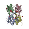
| Title | Structure of Mutant Pyrrolidone Carboxyl Peptidase (E192A) from a Hyperthermophile, Pyrococcus furiosus | ||||||
 Components Components | Pyrrolidone-carboxylate peptidase | ||||||
 Keywords Keywords | HYDROLASE / Stability of Protein | ||||||
| Function / homology |  Function and homology information Function and homology informationpyroglutamyl-peptidase I / pyroglutamyl-peptidase activity / proteolysis / cytosol Similarity search - Function | ||||||
| Biological species |   Pyrococcus furiosus (archaea) Pyrococcus furiosus (archaea) | ||||||
| Method |  X-RAY DIFFRACTION / SYNCHROTRON / MOLECULAR REPLACEMENT / Resolution: 2 Å X-RAY DIFFRACTION / SYNCHROTRON / MOLECULAR REPLACEMENT / Resolution: 2 Å | ||||||
 Authors Authors | Kaushik, J.K. / Yamagata, Y. / Ogasahara, K. / Yutani, K. | ||||||
 Citation Citation | Journal: Biochemistry / Year: 2006 Title: Completely buried, non-ion-paired glutamic acid contributes favorably to the conformational stability of pyrrolidone carboxyl peptidases from hyperthermophiles. Authors: Kaushik, J.K. / Iimura, S. / Ogasahara, K. / Yamagata, Y. / Segawa, S. / Yutani, K. #1: Journal: J.Biochem.(Tokyo) / Year: 2001Title: X-ray crystalline structures of pyrrolidone carboxyl peptidase from a hyperthermophile, Pyrococcus furiosus, and its cys-free mutant Authors: Tanaka, H. / Chinami, M. / Mizushima, T. / Ogasahara, K. / Ota, M. / Tsukihara, T. / Yutani, K. | ||||||
| History |
|
- Structure visualization
| Structure viewer | Molecule: MolmilJmol/JSmol |
|---|
- Downloads & links
-Download
| PDBx/mmCIF format | 1x10.cif.gz | 179 KB | Display | PDBx/mmCIF format |
|---|---|---|---|---|
| PDB format | pdb1x10.ent.gz | 143.3 KB | Display | PDB format |
| PDBx/mmJSON format | 1x10.json.gz | Tree view | PDBx/mmJSON format | |
| Others |  Other downloads Other downloads |
-Validation report
| Arichive directory | https://data.pdbj.org/pub/pdb/validation_reports/x1/1x10ftp://data.pdbj.org/pub/pdb/validation_reports/x1/1x10 | HTTPS FTP |
|---|
-Related structure data
| Related structure data | 1x12C  1z8tC  1z8wC 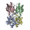 1z8xC 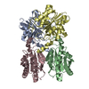 1ioiS S: Starting model for refinement C: citing same article ( |
|---|---|
| Similar structure data |


-Links
 PDBj
PDBj- Assembly
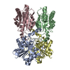
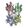
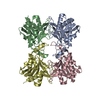
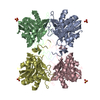
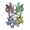
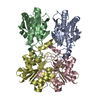
| Deposited unit |
| ||||||||
|---|---|---|---|---|---|---|---|---|---|
| 1 |
| ||||||||
| Unit cell |
|
-Components
| #1: Protein | Mass: 22761.619 Da / Num. of mol.: 4 / Mutation: C142S, C188S, E192A Source method: isolated from a genetically manipulated source Source: (gene. exp.) Pyrococcus furiosus (archaea) / Production host:  #2: Water | ChemComp-HOH / |  Mass: 18.015 Da / Num. of mol.: 724 / Source method: isolated from a natural source / Formula: H2O Mass: 18.015 Da / Num. of mol.: 724 / Source method: isolated from a natural source / Formula: H2O |
|---|
-Experimental details
-Experiment
| Experiment | Method: X-RAY DIFFRACTION / Number of used crystals: 1 |
|---|
- Sample preparation
| Crystal | Density Matthews: 2.54 Å3/Da / Density % sol: 51.52 % |
|---|---|
| Crystal grow | Temperature: 298 K / Method: vapor diffusion, hanging drop / pH: 4.6 Details: PEG4000, pH 4.6, VAPOR DIFFUSION, HANGING DROP, temperature 298K |
-Data collection
| Diffraction | Mean temperature: 100 K |
|---|---|
| Diffraction source | Source: SYNCHROTRON / Site: SPring-8  / Beamline: BL40B2 / Wavelength: 1 Å / Beamline: BL40B2 / Wavelength: 1 Å |
| Detector | Type: ADSC QUANTUM 4 / Detector: CCD / Date: Dec 15, 2000 |
| Radiation | Protocol: SINGLE WAVELENGTH / Monochromatic (M) / Laue (L): M / Scattering type: x-ray |
| Radiation wavelength | Wavelength: 1 Å / Relative weight: 1 |
| Reflection | Resolution: 2→50 Å / Num. all: 62100 / Num. obs: 62100 / % possible obs: 96.4 % / Observed criterion σ(F): 0 / Observed criterion σ(I): 0 / Redundancy: 5.1 % / Rmerge(I) obs: 0.048 / Net I/σ(I): 33 |
| Reflection shell | Resolution: 2→2.03 Å / Rmerge(I) obs: 0.083 / Mean I/σ(I) obs: 15.2 / % possible all: 86 |
- Processing
| Software |
| |||||||||||||||||||||||||
|---|---|---|---|---|---|---|---|---|---|---|---|---|---|---|---|---|---|---|---|---|---|---|---|---|---|---|
| Refinement | Method to determine structure: MOLECULAR REPLACEMENT Starting model: 1IOI Resolution: 2→20 Å / σ(F): 2 / Stereochemistry target values: Engh & Huber
| |||||||||||||||||||||||||
| Refinement step | Cycle: LAST / Resolution: 2→20 Å
|
