 Movie
Movie Controller

[English] 日本語
 Yorodumi
- PDB-1ioi: x-ray crystalline structures of pyrrolidone carboxyl peptidase fr... -
+ Open data

- Basic information
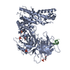
| Entry | Database: PDB / ID: 1ioi | ||||||
|---|---|---|---|---|---|---|---|
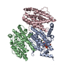
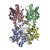
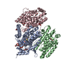
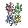
| Title | x-ray crystalline structures of pyrrolidone carboxyl peptidase from a hyperthermophile, pyrococcus furiosus, and its cys-free mutant | ||||||
 Components Components | PYRROLIDONE CARBOXYL PEPTIDASE | ||||||
 Keywords Keywords | HYDROLASE / PGP-I / PYROGLUTAMYL-PEPTIDASE I / PCP / protease / Pyrococcus furiosus / Archaea | ||||||
| Function / homology |  Function and homology information Function and homology informationpyroglutamyl-peptidase I / pyroglutamyl-peptidase activity / proteolysis / cytosol Similarity search - Function | ||||||
| Biological species |   Pyrococcus furiosus (archaea) Pyrococcus furiosus (archaea) | ||||||
| Method |  X-RAY DIFFRACTION / MOLECULAR REPLACEMENT / Resolution: 2.7 Å X-RAY DIFFRACTION / MOLECULAR REPLACEMENT / Resolution: 2.7 Å | ||||||
 Authors Authors | Tanaka, H. / Chinami, M. / Ota, M. / Tsukihara, T. / Yutani, K. | ||||||
 Citation Citation | Journal: J.Biochem. / Year: 2001 Title: X-ray crystalline structures of pyrrolidone carboxyl peptidase from a hyperthermophile, Pyrococcus furiosus, and its cys-free mutant. Authors: Tanaka, H. / Chinami, M. / Mizushima, T. / Ogasahara, K. / Ota, M. / Tsukihara, T. / Yutani, K. | ||||||
| History |
|
- Structure visualization
| Structure viewer | Molecule: MolmilJmol/JSmol |
|---|
- Downloads & links
-Download
| PDBx/mmCIF format | 1ioi.cif.gz | 166.8 KB | Display | PDBx/mmCIF format |
|---|---|---|---|---|
| PDB format | pdb1ioi.ent.gz | 134 KB | Display | PDB format |
| PDBx/mmJSON format | 1ioi.json.gz | Tree view | PDBx/mmJSON format | |
| Others |  Other downloads Other downloads |
-Validation report
| Arichive directory | https://data.pdbj.org/pub/pdb/validation_reports/io/1ioiftp://data.pdbj.org/pub/pdb/validation_reports/io/1ioi | HTTPS FTP |
|---|
-Related structure data
| Related structure data |  1iofC 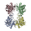 1augS C: citing same article ( S: Starting model for refinement |
|---|---|
| Similar structure data |

-Links
 PDBj
PDBj- Assembly
| Deposited unit | 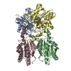
| ||||||||
|---|---|---|---|---|---|---|---|---|---|
| 1 |
| ||||||||
| Unit cell |
| ||||||||
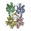
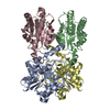
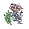
| Details | The biological assembly is a tetramer. |
-Components
| #1: Protein | Mass: 22819.654 Da / Num. of mol.: 4 / Mutation: C142S/C188S Source method: isolated from a genetically manipulated source Source: (gene. exp.) Pyrococcus furiosus (archaea) / Plasmid: PPCP3022 / Production host:  #2: Water | ChemComp-HOH / |  Mass: 18.015 Da / Num. of mol.: 124 / Source method: isolated from a natural source / Formula: H2O Mass: 18.015 Da / Num. of mol.: 124 / Source method: isolated from a natural source / Formula: H2O |
|---|
-Experimental details
-Experiment
| Experiment | Method: X-RAY DIFFRACTION / Number of used crystals: 2 |
|---|
- Sample preparation
| Crystal | Density Matthews: 2.98 Å3/Da / Density % sol: 58.79 % | ||||||||||||||||||||||||||||||
|---|---|---|---|---|---|---|---|---|---|---|---|---|---|---|---|---|---|---|---|---|---|---|---|---|---|---|---|---|---|---|---|
| Crystal grow | Temperature: 298 K / Method: vapor diffusion, hanging drop / pH: 4.6 Details: ammonium sulfate, MPD, sodium acetate, EDTA, DTE, pH 4.6, VAPOR DIFFUSION, HANGING DROP, temperature 298.0K | ||||||||||||||||||||||||||||||
| Crystal grow | *PLUS Temperature: 25 ℃ | ||||||||||||||||||||||||||||||
| Components of the solutions | *PLUS
|
-Data collection
| Diffraction | Mean temperature: 298 K |
|---|---|
| Diffraction source | Source: ROTATING ANODE / Type: RIGAKU RU200 / Wavelength: 1.5418 Å |
| Detector | Type: MAC Science DIP-2030 / Detector: IMAGE PLATE / Date: Oct 24, 1996 / Details: nickel-filtered double-mirror |
| Radiation | Monochromator: nickel-filtered double-mirror / Protocol: SINGLE WAVELENGTH / Monochromatic (M) / Laue (L): M / Scattering type: x-ray |
| Radiation wavelength | Wavelength: 1.5418 Å / Relative weight: 1 |
| Reflection | Resolution: 2.5→200 Å / Num. all: 281217 / Num. obs: 118965 / % possible obs: 89.9 % / Observed criterion σ(F): 1 / Observed criterion σ(I): 1 / Redundancy: 3.6 % / Rmerge(I) obs: 0.13 / Net I/σ(I): 5 |
| Reflection shell | Resolution: 2.5→2.61 Å / Redundancy: 1.9 % / Rmerge(I) obs: 0.211 / Mean I/σ(I) obs: 2.8 / Num. unique all: 3349 / % possible all: 72 |
| Reflection | *PLUS Num. obs: 33343 |
| Reflection shell | *PLUS % possible obs: 72 % |
- Processing
| Software |
| ||||||||||||||||||||||||||||||
|---|---|---|---|---|---|---|---|---|---|---|---|---|---|---|---|---|---|---|---|---|---|---|---|---|---|---|---|---|---|---|---|
| Refinement | Method to determine structure: MOLECULAR REPLACEMENT Starting model: ENTRY 1AUG Resolution: 2.7→10 Å / σ(F): 2 / σ(I): 2 / Stereochemistry target values: Engh & Huber
| ||||||||||||||||||||||||||||||
| Refinement step | Cycle: LAST / Resolution: 2.7→10 Å
| ||||||||||||||||||||||||||||||
| Software | *PLUS Name: 'X-PLOR' / Classification: refinement | ||||||||||||||||||||||||||||||
| Refine LS restraints | *PLUS
|
