 Movie
Movie Controller


+ Open data

- Basic information
| Entry | Database: PDB / ID: 1wz3 | ||||||
|---|---|---|---|---|---|---|---|
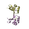
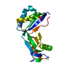
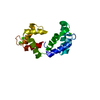
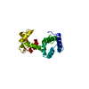
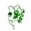
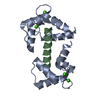
| Title | The crystal structure of plant ATG12 | ||||||
 Components Components | autophagy 12b | ||||||
 Keywords Keywords | PLANT PROTEIN / ubiquitin-fold | ||||||
| Function / homology |  Function and homology information Function and homology information | ||||||
| Biological species |  | ||||||
| Method |  X-RAY DIFFRACTION / MIRAS / Resolution: 1.8 Å X-RAY DIFFRACTION / MIRAS / Resolution: 1.8 Å | ||||||
 Authors Authors | Suzuki, N.N. / Yoshimoto, K. / Fujioka, Y. / Ohsumi, Y. / Inagaki, F. | ||||||
 Citation Citation | Journal: Autophagy / Year: 2005 Title: The crystal structure of plant ATG12 and its biological implication in autophagy. Authors: Suzuki, N.N. / Yoshimoto, K. / Fujioka, Y. / Ohsumi, Y. / Inagaki, F. | ||||||
| History |
|
- Structure visualization
| Structure viewer | Molecule: MolmilJmol/JSmol |
|---|
- Downloads & links
-Download
| PDBx/mmCIF format | 1wz3.cif.gz | 47.3 KB | Display | PDBx/mmCIF format |
|---|---|---|---|---|
| PDB format | pdb1wz3.ent.gz | 33.2 KB | Display | PDB format |
| PDBx/mmJSON format | 1wz3.json.gz | Tree view | PDBx/mmJSON format | |
| Others |  Other downloads Other downloads |
-Validation report
| Summary document | 1wz3_validation.pdf.gz | 435.2 KB | Display | wwPDB validaton report |
|---|---|---|---|---|
| Full document | 1wz3_full_validation.pdf.gz | 436.6 KB | Display | |
| Data in XML | 1wz3_validation.xml.gz | 10.3 KB | Display | |
| Data in CIF | 1wz3_validation.cif.gz | 13.8 KB | Display | |
| Arichive directory | https://data.pdbj.org/pub/pdb/validation_reports/wz/1wz3ftp://data.pdbj.org/pub/pdb/validation_reports/wz/1wz3 | HTTPS FTP |
-Related structure data
| Similar structure data |
|---|

-Links
 PDBj
PDBj- Assembly
| Deposited unit | 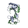
| ||||||||
|---|---|---|---|---|---|---|---|---|---|
| 1 |
| ||||||||
| Unit cell |
|
-Components
| #1: Protein | Mass: 10522.900 Da / Num. of mol.: 2 Source method: isolated from a genetically manipulated source Source: (gene. exp.)  #2: Water | ChemComp-HOH / |  Mass: 18.015 Da / Num. of mol.: 156 / Source method: isolated from a natural source / Formula: H2O Mass: 18.015 Da / Num. of mol.: 156 / Source method: isolated from a natural source / Formula: H2O |
|---|
-Experimental details
-Experiment
| Experiment | Method: X-RAY DIFFRACTION / Number of used crystals: 1 |
|---|
- Sample preparation
| Crystal | Density Matthews: 2.11 Å3/Da / Density % sol: 41.65 % |
|---|---|
| Crystal grow | Temperature: 293 K / Method: vapor diffusion, sitting drop / pH: 7.4 Details: PEG8000, HEPES, pH 7.4, VAPOR DIFFUSION, SITTING DROP, temperature 293K |
-Data collection
| Diffraction | Mean temperature: 100 K |
|---|---|
| Diffraction source | Source: ROTATING ANODE / Type: RIGAKU / Wavelength: 1.5418 Å |
| Detector | Type: RIGAKU RAXIS / Detector: IMAGE PLATE |
| Radiation | Protocol: SINGLE WAVELENGTH / Monochromatic (M) / Laue (L): M / Scattering type: x-ray |
| Radiation wavelength | Wavelength: 1.5418 Å / Relative weight: 1 |
| Reflection | Resolution: 1.8→50 Å / Num. all: 16031 / Num. obs: 16031 / % possible obs: 97.3 % / Observed criterion σ(F): 0 / Observed criterion σ(I): -3 / Biso Wilson estimate: 21.8 Å2 / Rmerge(I) obs: 0.04 / Net I/σ(I): 20.8 |
| Reflection shell | Resolution: 1.8→1.86 Å / Rmerge(I) obs: 0.275 / % possible all: 80.5 |
- Processing
| Software |
| |||||||||||||||||||||||||
|---|---|---|---|---|---|---|---|---|---|---|---|---|---|---|---|---|---|---|---|---|---|---|---|---|---|---|
| Refinement | Method to determine structure: MIRAS / Resolution: 1.8→28.92 Å / Rfactor Rfree error: 0.006 / Data cutoff high absF: 564936.56 / Data cutoff low absF: 0 / Isotropic thermal model: RESTRAINED / Cross valid method: THROUGHOUT / σ(F): 0 / Stereochemistry target values: Engh & Huber
| |||||||||||||||||||||||||
| Solvent computation | Solvent model: FLAT MODEL / Bsol: 64.9019 Å2 / ksol: 0.403451 e/Å3 | |||||||||||||||||||||||||
| Displacement parameters | Biso mean: 28.6 Å2
| |||||||||||||||||||||||||
| Refine analyze |
| |||||||||||||||||||||||||
| Refinement step | Cycle: LAST / Resolution: 1.8→28.92 Å
| |||||||||||||||||||||||||
| Refine LS restraints |
| |||||||||||||||||||||||||
| LS refinement shell | Resolution: 1.8→1.91 Å / Rfactor Rfree error: 0.029 / Total num. of bins used: 6
| |||||||||||||||||||||||||
| Xplor file |
|
