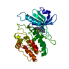
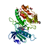
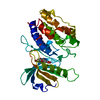
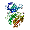
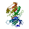
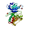
- PDB-1wvw: Crystal structures of kinase domain of DAP kinase in complex with... -
+
データを開く
IDまたはキーワード:
読み込み中...
-
基本情報
登録情報
データベース: PDB / ID: 1wvw
タイトル
Crystal structures of kinase domain of DAP kinase in complex with small molecular inhibitors
要素
Death-associated protein kinase 1
キーワード
TRANSFERASE / protein kinase / apoptosis
機能・相同性
機能・相同性情報
cellular response to hydroperoxide / regulation of response to tumor cell / positive regulation of autophagic cell death / DAPK1-calmodulin complex / defense response to tumor cell / Caspase activation via Dependence Receptors in the absence of ligand / regulation of NMDA receptor activity / syntaxin-1 binding / calcium/calmodulin-dependent protein kinase activity / positive regulation of cysteine-type endopeptidase activity involved in apoptotic process ...cellular response to hydroperoxide / regulation of response to tumor cell / positive regulation of autophagic cell death / DAPK1-calmodulin complex / defense response to tumor cell / Caspase activation via Dependence Receptors in the absence of ligand / regulation of NMDA receptor activity / syntaxin-1 binding / calcium/calmodulin-dependent protein kinase activity / positive regulation of cysteine-type endopeptidase activity involved in apoptotic process / extrinsic apoptotic signaling pathway via death domain receptors / positive regulation of autophagy / regulation of autophagy / apoptotic signaling pathway / cellular response to type II interferon / actin cytoskeleton / regulation of apoptotic process / protein autophosphorylation / postsynaptic density / negative regulation of translation / non-specific serine/threonine protein kinase / calmodulin binding / protein kinase activity / intracellular signal transduction / positive regulation of apoptotic process / protein phosphorylation / protein serine kinase activity / protein serine/threonine kinase activity / glutamatergic synapse / apoptotic process / GTP binding / negative regulation of apoptotic process / ATP binding / identical protein binding / nucleus / plasma membrane / cytoplasm 類似検索 - 分子機能
Death-associated protein kinase 1 / Roc domain profile. / Roc domain / Death domain profile. / DEATH domain, found in proteins involved in cell death (apoptosis). / Death domain / Death domain / Death-like domain superfamily / Ankyrin repeat / Ankyrin repeats (3 copies) ...Death-associated protein kinase 1 / Roc domain profile. / Roc domain / Death domain profile. / DEATH domain, found in proteins involved in cell death (apoptosis). / Death domain / Death domain / Death-like domain superfamily / Ankyrin repeat / Ankyrin repeats (3 copies) / Ankyrin repeat profile. / Ankyrin repeat region circular profile. / ankyrin repeats / Ankyrin repeat / Ankyrin repeat-containing domain superfamily / Transferase(Phosphotransferase) domain 1 / Transferase(Phosphotransferase); domain 1 / Phosphorylase Kinase; domain 1 / Phosphorylase Kinase; domain 1 / Serine/threonine-protein kinase, active site / Serine/Threonine protein kinases active-site signature. / Protein kinase domain / Serine/Threonine protein kinases, catalytic domain / Protein kinase, ATP binding site / Protein kinases ATP-binding region signature. / Protein kinase domain profile. / Protein kinase domain / Protein kinase-like domain superfamily / P-loop containing nucleoside triphosphate hydrolase / 2-Layer Sandwich / Orthogonal Bundle / Mainly Alpha / Alpha Beta 類似検索 - ドメイン・相同性
 ムービー
ムービー コントローラー


 データを開く
 基本情報
基本情報 要素
要素 キーワード
キーワード 機能・相同性情報
 Homo sapiens (ヒト)
 X線回折 /
  データ登録者
 引用
引用 構造の表示
構造の表示 ダウンロードとリンク
 その他のダウンロード


 PDBj
PDBj


 集合体
集合体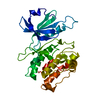

 試料調製
試料調製 / ビームライン: BL40B2 / 波長: 0.9 Å
 解析
解析