 Movie
Movie Controller


+ Open data

- Basic information
| Entry | Database: PDB / ID: 1wq8 | |||||||||
|---|---|---|---|---|---|---|---|---|---|---|
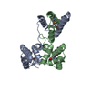
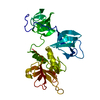
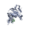
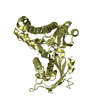
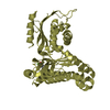
| Title | Crystal structure of Vammin, a VEGF-F from a snake venom | |||||||||
 Components Components | Vascular endothelial growth factor toxin | |||||||||
 Keywords Keywords | TOXIN / snake venom / Vascular endothelial growth factor / VEGF / VEGF-F | |||||||||
| Function / homology |  Function and homology information Function and homology informationvascular endothelial growth factor receptor binding / positive regulation of mast cell chemotaxis / induction of positive chemotaxis / sprouting angiogenesis / vascular endothelial growth factor signaling pathway / chemoattractant activity / vascular endothelial growth factor receptor signaling pathway / positive regulation of endothelial cell proliferation / growth factor activity / positive regulation of angiogenesis ...vascular endothelial growth factor receptor binding / positive regulation of mast cell chemotaxis / induction of positive chemotaxis / sprouting angiogenesis / vascular endothelial growth factor signaling pathway / chemoattractant activity / vascular endothelial growth factor receptor signaling pathway / positive regulation of endothelial cell proliferation / growth factor activity / positive regulation of angiogenesis / toxin activity / response to hypoxia / extracellular space / membrane Similarity search - Function | |||||||||
| Biological species |  Vipera aspis aspis (snake) Vipera aspis aspis (snake) | |||||||||
| Method |  X-RAY DIFFRACTION / SYNCHROTRON / MOLECULAR REPLACEMENT / Resolution: 1.9 Å X-RAY DIFFRACTION / SYNCHROTRON / MOLECULAR REPLACEMENT / Resolution: 1.9 Å | |||||||||
 Authors Authors | Suto, K. / Yamazaki, Y. / Morita, T. / Mizuno, H. | |||||||||
 Citation Citation | Journal: J.Biol.Chem. / Year: 2005 Title: Crystal structures of novel vascular endothelial growth factors (VEGF) from snake venoms: insight into selective VEGF binding to kinase insert domain-containing receptor but not to fms-like tyrosine kinase-1. Authors: Suto, K. / Yamazaki, Y. / Morita, T. / Mizuno, H. #1: Journal: J.Biol.Chem. / Year: 2003 Title: Snake venom vascular endothelial growth factors (VEGFs) exhibit potent activity through their specific recognition of KDR (VEGF receptor 2) Authors: Yamazaki, Y. / Takani, K. / Atoda, H. / Morita, T. | |||||||||
| History |
|
- Structure visualization
| Structure viewer | Molecule: MolmilJmol/JSmol |
|---|
- Downloads & links
-Download
| PDBx/mmCIF format | 1wq8.cif.gz | 37.9 KB | Display | PDBx/mmCIF format |
|---|---|---|---|---|
| PDB format | pdb1wq8.ent.gz | 25.2 KB | Display | PDB format |
| PDBx/mmJSON format | 1wq8.json.gz | Tree view | PDBx/mmJSON format | |
| Others |  Other downloads Other downloads |
-Validation report
| Summary document | 1wq8_validation.pdf.gz | 442.9 KB | Display | wwPDB validaton report |
|---|---|---|---|---|
| Full document | 1wq8_full_validation.pdf.gz | 443.5 KB | Display | |
| Data in XML | 1wq8_validation.xml.gz | 8.5 KB | Display | |
| Data in CIF | 1wq8_validation.cif.gz | 11.7 KB | Display | |
| Arichive directory | https://data.pdbj.org/pub/pdb/validation_reports/wq/1wq8ftp://data.pdbj.org/pub/pdb/validation_reports/wq/1wq8 | HTTPS FTP |
-Related structure data


-Links
 PDBj
PDBj

- Assembly
| Deposited unit | 
| ||||||||||||
|---|---|---|---|---|---|---|---|---|---|---|---|---|---|
| 1 | 
| ||||||||||||
| 2 | x 8
| ||||||||||||
| Unit cell |
| ||||||||||||
| Components on special symmetry positions |
| ||||||||||||
| Details | The second part of the biological assembly is generated by the two fold axis:y, x, -z |
-Components
| #1: Protein | Mass: 12575.499 Da / Num. of mol.: 1 / Source method: isolated from a natural source / Source: (natural) Vipera aspis aspis (snake) / Secretion: venom / Species: Vipera aspis / Strain: aspis / References: UniProt: P83942, UniProt: P67863*PLUS |
|---|---|
| #2: Chemical | ChemComp-TRS /   Mass: 122.143 Da / Num. of mol.: 1 / Source method: obtained synthetically / Formula: C4H12NO3 / Comment: pH buffer*YM Mass: 122.143 Da / Num. of mol.: 1 / Source method: obtained synthetically / Formula: C4H12NO3 / Comment: pH buffer*YM |
| #3: Water | ChemComp-HOH /  Mass: 18.015 Da / Num. of mol.: 172 / Source method: isolated from a natural source / Formula: H2O Mass: 18.015 Da / Num. of mol.: 172 / Source method: isolated from a natural source / Formula: H2O |
| Has protein modification | Y |
-Experimental details
-Experiment
| Experiment | Method: X-RAY DIFFRACTION / Number of used crystals: 1 |
|---|
- Sample preparation
| Crystal | Density Matthews: 3.2 Å3/Da / Density % sol: 62 % |
|---|---|
| Crystal grow | Temperature: 293 K / Method: vapor diffusion, sitting drop / pH: 9 Details: PEG 6000, Tris-HCl, glycerol, pH 9.0, VAPOR DIFFUSION, SITTING DROP, temperature 293K |
-Data collection
| Diffraction | Mean temperature: 100 K |
|---|---|
| Diffraction source | Source: SYNCHROTRON / Site: Photon Factory  / Beamline: AR-NW12A / Wavelength: 1 Å / Beamline: AR-NW12A / Wavelength: 1 Å |
| Detector | Type: ADSC QUANTUM 210 / Detector: CCD / Date: Jun 13, 2003 |
| Radiation | Monochromator: SILICON / Protocol: SINGLE WAVELENGTH / Monochromatic (M) / Laue (L): M / Scattering type: x-ray |
| Radiation wavelength | Wavelength: 1 Å / Relative weight: 1 |
| Reflection | Resolution: 1.9→50 Å / Num. all: 13068 / Num. obs: 12903 / % possible obs: 99.7 % / Observed criterion σ(F): 0 / Observed criterion σ(I): -3 / Biso Wilson estimate: 16.7 Å2 / Rmerge(I) obs: 0.048 |
| Reflection shell | Resolution: 1.9→1.97 Å / Rmerge(I) obs: 0.218 / % possible all: 100 |
- Processing
| Software |
| ||||||||||||||||||||||||||||||||||||||||||||||||||||||||||||||||||||||||||||||||
|---|---|---|---|---|---|---|---|---|---|---|---|---|---|---|---|---|---|---|---|---|---|---|---|---|---|---|---|---|---|---|---|---|---|---|---|---|---|---|---|---|---|---|---|---|---|---|---|---|---|---|---|---|---|---|---|---|---|---|---|---|---|---|---|---|---|---|---|---|---|---|---|---|---|---|---|---|---|---|---|---|---|
| Refinement | Method to determine structure: MOLECULAR REPLACEMENT Starting model: VR-1 Resolution: 1.9→23 Å / Rfactor Rfree error: 0.009 / Data cutoff high absF: 1706273.81 / Data cutoff low absF: 0 / Isotropic thermal model: RESTRAINED / Cross valid method: THROUGHOUT / σ(F): 0 / Stereochemistry target values: Engh & Huber
| ||||||||||||||||||||||||||||||||||||||||||||||||||||||||||||||||||||||||||||||||
| Solvent computation | Solvent model: FLAT MODEL / Bsol: 51.5954 Å2 / ksol: 0.377209 e/Å3 | ||||||||||||||||||||||||||||||||||||||||||||||||||||||||||||||||||||||||||||||||
| Displacement parameters | Biso mean: 29.1 Å2
| ||||||||||||||||||||||||||||||||||||||||||||||||||||||||||||||||||||||||||||||||
| Refine analyze |
| ||||||||||||||||||||||||||||||||||||||||||||||||||||||||||||||||||||||||||||||||
| Refinement step | Cycle: LAST / Resolution: 1.9→23 Å
| ||||||||||||||||||||||||||||||||||||||||||||||||||||||||||||||||||||||||||||||||
| Refine LS restraints |
| ||||||||||||||||||||||||||||||||||||||||||||||||||||||||||||||||||||||||||||||||
| LS refinement shell | Resolution: 1.9→2.02 Å / Rfactor Rfree error: 0.023 / Total num. of bins used: 6
| ||||||||||||||||||||||||||||||||||||||||||||||||||||||||||||||||||||||||||||||||
| Xplor file |
|
