| 登録情報 | データベース: PDB / ID: 1wmh
|
|---|
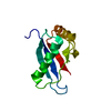
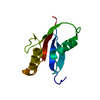
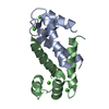
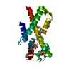
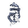
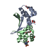
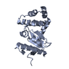
| タイトル | Crystal structure of a PB1 domain complex of Protein kinase c iota and Par6 alpha |
|---|
 要素 要素 | - Partitioning defective-6 homolog alpha
- Protein kinase C, iota type
|
|---|
 キーワード キーワード | Transferase/CELL CYCLE / KINASE / PB1 DOMAIN / OPCA MOTIF / aPKC / Par6 / cell polarity / Transferase-CELL CYCLE COMPLEX |
|---|
| 機能・相同性 |  機能・相同性情報 機能・相同性情報
regulation of cellular localization / diacylglycerol-dependent, calcium-independent serine/threonine kinase activity / Golgi vesicle budding / PAR polarity complex / Tight junction interactions / cell-cell junction maintenance / protein kinase C / establishment of apical/basal cell polarity / diacylglycerol-dependent serine/threonine kinase activity / negative regulation of glial cell apoptotic process ...regulation of cellular localization / diacylglycerol-dependent, calcium-independent serine/threonine kinase activity / Golgi vesicle budding / PAR polarity complex / Tight junction interactions / cell-cell junction maintenance / protein kinase C / establishment of apical/basal cell polarity / diacylglycerol-dependent serine/threonine kinase activity / negative regulation of glial cell apoptotic process / eye photoreceptor cell development / positive regulation of protein localization to centrosome / GTP-dependent protein binding / Schmidt-Lanterman incisure / establishment or maintenance of epithelial cell apical/basal polarity / membrane organization / cellular response to chemical stress / cell-cell junction organization / centrosome cycle / protein targeting to membrane / tight junction / RHOV GTPase cycle / positive regulation of Notch signaling pathway / establishment of cell polarity / establishment or maintenance of cell polarity / cell leading edge / RHOU GTPase cycle / brush border / CDC42 GTPase cycle / positive regulation of endothelial cell apoptotic process / positive regulation of glial cell proliferation / viral process / bicellular tight junction / regulation of postsynaptic membrane neurotransmitter receptor levels / intercellular bridge / vesicle-mediated transport / ruffle / RAC1 GTPase cycle / cytoskeleton organization / secretion / p75NTR recruits signalling complexes / response to interleukin-1 / actin filament organization / TGF-beta receptor signaling in EMT (epithelial to mesenchymal transition) / positive regulation of D-glucose import / positive regulation of protein secretion / protein localization to plasma membrane / Asymmetric localization of PCP proteins / positive regulation of protein localization to plasma membrane / positive regulation of NF-kappaB transcription factor activity / positive regulation of neuron projection development / phospholipid binding / small GTPase binding / Pre-NOTCH Transcription and Translation / centriolar satellite / Schaffer collateral - CA1 synapse / cellular response to insulin stimulus / KEAP1-NFE2L2 pathway / cell migration / microtubule cytoskeleton / cell cortex / negative regulation of neuron apoptotic process / protein phosphorylation / protein kinase activity / endosome / intracellular signal transduction / cilium / apical plasma membrane / Golgi membrane / cell division / protein serine kinase activity / intracellular membrane-bounded organelle / protein serine/threonine kinase activity / centrosome / negative regulation of apoptotic process / glutamatergic synapse / extracellular exosome / zinc ion binding / nucleoplasm / ATP binding / nucleus / plasma membrane / cytosol類似検索 - 分子機能 Partitioning defective protein 6, PB1 domain / : / Atypical protein kinase C iota type, catalytic domain / Protein kinase C / Protein kinase C, PB1 domain / PB1 domain / PB1 domain / PB1 domain / : / PB1 domain profile. ...Partitioning defective protein 6, PB1 domain / : / Atypical protein kinase C iota type, catalytic domain / Protein kinase C / Protein kinase C, PB1 domain / PB1 domain / PB1 domain / PB1 domain / : / PB1 domain profile. / Protein kinase, C-terminal / Protein kinase C terminal domain / Diacylglycerol/phorbol-ester binding / Phorbol esters/diacylglycerol binding domain (C1 domain) / Zinc finger phorbol-ester/DAG-type signature. / Zinc finger phorbol-ester/DAG-type profile. / Protein kinase C conserved region 1 (C1) domains (Cysteine-rich domains) / Protein kinase C-like, phorbol ester/diacylglycerol-binding domain / C1-like domain superfamily / Extension to Ser/Thr-type protein kinases / AGC-kinase, C-terminal / AGC-kinase C-terminal domain profile. / Phosphatidylinositol 3-kinase Catalytic Subunit; Chain A, domain 1 / PDZ domain / PDZ domain profile. / Domain present in PSD-95, Dlg, and ZO-1/2. / PDZ domain / PDZ superfamily / Ubiquitin-like (UB roll) / Serine/threonine-protein kinase, active site / Serine/Threonine protein kinases active-site signature. / Protein kinase domain / Serine/Threonine protein kinases, catalytic domain / Roll / Protein kinase, ATP binding site / Protein kinases ATP-binding region signature. / Protein kinase domain profile. / Protein kinase domain / Protein kinase-like domain superfamily / Alpha Beta類似検索 - ドメイン・相同性 Protein kinase C iota type / Partitioning defective 6 homolog alpha類似検索 - 構成要素 |
|---|
| 生物種 |  Homo sapiens (ヒト) Homo sapiens (ヒト) |
|---|
| 手法 |  X線回折 / シンクロトロン / 多波長異常分散 / 解像度: 1.5 Å X線回折 / シンクロトロン / 多波長異常分散 / 解像度: 1.5 Å |
|---|
 データ登録者 データ登録者 | Hirano, Y. / Yoshinaga, S. / Suzuki, N.N. / Horiuchi, M. / Kohjima, M. / Takeya, R. / Sumimoto, H. / Inagaki, F. |
|---|
 引用 引用 | ジャーナル: J.Biol.Chem. / 年: 2005
タイトル: Structure of a Cell Polarity Regulator, a Complex between Atypical PKC and Par6 PB1 Domains
著者: Hirano, Y. / Yoshinaga, S. / Takeya, R. / Suzuki, N.N. / Horiuchi, M. / Kohjima, M. / Sumimoto, H. / Inagaki, F. |
|---|
| 履歴 | | 登録 | 2004年7月9日 | 登録サイト: PDBJ / 処理サイト: PDBJ |
|---|
| 改定 1.0 | 2004年12月7日 | Provider: repository / タイプ: Initial release |
|---|
| 改定 1.1 | 2008年4月30日 | Group: Version format compliance |
|---|
| 改定 1.2 | 2011年7月13日 | Group: Version format compliance |
|---|
| 改定 2.0 | 2024年3月13日 | Group: Atomic model / Data collection / Database references
カテゴリ: atom_site / chem_comp_atom ...atom_site / chem_comp_atom / chem_comp_bond / database_2 / struct_ref_seq_dif
Item: _atom_site.occupancy / _database_2.pdbx_DOI ..._atom_site.occupancy / _database_2.pdbx_DOI / _database_2.pdbx_database_accession / _struct_ref_seq_dif.details |
|---|
|
|---|
 ムービー
ムービー コントローラー


 データを開く
 基本情報
基本情報 構造の表示
構造の表示 ダウンロードとリンク
 その他のダウンロード
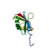


 PDBj
PDBj


 集合体
集合体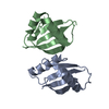

 分子量: 18.015 Da / 分子数: 188 / 由来タイプ: 天然 / 式: H2O
 試料調製
試料調製 / ビームライン: BL41XU / 波長: 0.9791, 0.9793, 0.9795, 0.9843
 解析
解析