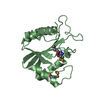
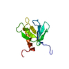
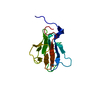
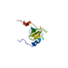
ERK5 cascade / negative regulation of chemokine (C-X-C motif) ligand 2 production / negative regulation of response to cytokine stimulus / negative regulation of heterotypic cell-cell adhesion / mitogen-activated protein kinase kinase / negative regulation of interleukin-8 production / negative regulation of cell migration involved in sprouting angiogenesis / MAP kinase kinase activity / negative regulation of smooth muscle cell apoptotic process / positive regulation of protein metabolic process ...ERK5 cascade / negative regulation of chemokine (C-X-C motif) ligand 2 production / negative regulation of response to cytokine stimulus / negative regulation of heterotypic cell-cell adhesion / mitogen-activated protein kinase kinase / negative regulation of interleukin-8 production / negative regulation of cell migration involved in sprouting angiogenesis / MAP kinase kinase activity / negative regulation of smooth muscle cell apoptotic process / positive regulation of protein metabolic process / negative regulation of extrinsic apoptotic signaling pathway in absence of ligand / insulin-like growth factor receptor signaling pathway / positive regulation of epithelial cell proliferation / negative regulation of canonical NF-kappaB signal transduction / cellular response to growth factor stimulus / spindle / MAPK cascade / heart development / positive regulation of cell growth / protein tyrosine kinase activity / protein kinase activity / protein serine kinase activity / protein serine/threonine kinase activity / negative regulation of transcription by RNA polymerase II / positive regulation of transcription by RNA polymerase II / ATP binding / metal ion binding / cytoplasm Similarity search - Function
Dual specificity mitogen-activated protein kinase kinase 5, PB1 domain / : / PB1 domain / PB1 domain / PB1 domain / : / PB1 domain profile. / Phosphatidylinositol 3-kinase Catalytic Subunit; Chain A, domain 1 / Ubiquitin-like (UB roll) / Serine/threonine-protein kinase, active site ...Dual specificity mitogen-activated protein kinase kinase 5, PB1 domain / : / PB1 domain / PB1 domain / PB1 domain / : / PB1 domain profile. / Phosphatidylinositol 3-kinase Catalytic Subunit; Chain A, domain 1 / Ubiquitin-like (UB roll) / Serine/threonine-protein kinase, active site / Serine/Threonine protein kinases active-site signature. / Protein kinase domain / Serine/Threonine protein kinases, catalytic domain / Roll / Protein kinase, ATP binding site / Protein kinases ATP-binding region signature. / Protein kinase domain profile. / Protein kinase domain / Protein kinase-like domain superfamily / Alpha Beta Similarity search - Domain/homology
Conformer selection criteria: structures with the least restraint violations, target function Conformers calculated total number: 100 / Conformers submitted total number: 20
+
About Yorodumi
-
News
-
Feb 9, 2022. New format data for meta-information of EMDB entries
New format data for meta-information of EMDB entries
Version 3 of the EMDB header file is now the official format.
The previous official version 1.9 will be removed from the archive.
In the structure databanks used in Yorodumi, some data are registered as the other names, "COVID-19 virus" and "2019-nCoV". Here are the details of the virus and the list of structure data.
Jan 31, 2019. EMDB accession codes are about to change! (news from PDBe EMDB page)
EMDB accession codes are about to change! (news from PDBe EMDB page)
The allocation of 4 digits for EMDB accession codes will soon come to an end. Whilst these codes will remain in use, new EMDB accession codes will include an additional digit and will expand incrementally as the available range of codes is exhausted. The current 4-digit format prefixed with “EMD-” (i.e. EMD-XXXX) will advance to a 5-digit format (i.e. EMD-XXXXX), and so on. It is currently estimated that the 4-digit codes will be depleted around Spring 2019, at which point the 5-digit format will come into force.
The EM Navigator/Yorodumi systems omit the EMD- prefix.
Related info.:Q: What is EMD? / ID/Accession-code notation in Yorodumi/EM Navigator
Yorodumi is a browser for structure data from EMDB, PDB, SASBDB, etc.
This page is also the successor to EM Navigator detail page, and also detail information page/front-end page for Omokage search.
The word "yorodu" (or yorozu) is an old Japanese word meaning "ten thousand". "mi" (miru) is to see.
Related info.:EMDB / PDB / SASBDB / Comparison of 3 databanks / Yorodumi Search / Aug 31, 2016. New EM Navigator & Yorodumi / Yorodumi Papers / Jmol/JSmol / Function and homology information / Changes in new EM Navigator and Yorodumi
 Movie
Movie Controller

 Yorodumi
 Open data
 Basic information
 Components
 Keywords
 Function and homology information
 Authors
 Citation
 Structure visualization
 Downloads & links
 Other downloads


 PDBj
PDBj


 Assembly
 Sample preparation
 Processing
 CYANA
CYANA