[English] 日本語
Yorodumi
- PDB-1wck: Crystal structure of the C-terminal domain of BclA, the major ant... -

+
Open data


ID or keywords:

Loading...

-
Basic information

Entry
Database: PDB / ID: 1wck
TitleCrystal structure of the C-terminal domain of BclA, the major antigen of the exosporium of the Bacillus anthracis spore.
ComponentsBCLA PROTEIN
KeywordsSTRUCTURAL PROTEIN / COLLAGEN-LIKE PROTEIN / BACTERIAL SURFACE ANTIGEN / JELLY-ROLL TOPOLOGY
Function / homology
Function and homology information


extracellular matrix structural constituent conferring tensile strength / extracellular matrix organization / extracellular space
Similarity search - Function
Leader peptide, exosporium / BclA, C-terminal domain / BclA C-terminal domain / Jelly Rolls - #40 / Tumour necrosis factor-like domain superfamily / Collagen triple helix repeat / Collagen triple helix repeat (20 copies) / Jelly Rolls / Sandwich / Mainly Beta
Similarity search - Domain/homology
CACODYLATE ION / BclA protein / BclA protein
Similarity search - Component
Biological speciesBACILLUS ANTHRACIS (anthrax bacterium)
MethodX-RAY DIFFRACTION / SYNCHROTRON / SIRAS / Resolution: 1.36 Å
AuthorsRety, S. / Salamitou, S. / Augusto, L.A. / Chaby, R. / Lehegarat, F. / Lewit-Bentley, A.
CitationJournal: J.Biol.Chem. / Year: 2005
Title: The Crystal Structure of the Bacillus Anthracis Spore Surface Protein Bcla Shows Remarkable Similarity to Mammalian Proteins.
Authors: Rety, S. / Salamitou, S. / Garcia-Verdugo, I. / Hulmes, D.J. / Lehegarat, F. / Chaby, R. / Lewit-Bentley, A.
History
DepositionNov 17, 2004Deposition site: PDBE / Processing site: PDBE
Revision 1.0Oct 25, 2005Provider: repository / Type: Initial release
Revision 1.1May 8, 2011Group: Version format compliance
Revision 1.2Jul 13, 2011Group: Version format compliance
Revision 1.3Mar 6, 2019Group: Data collection / Derived calculations ...Data collection / Derived calculations / Experimental preparation / Other
Category: exptl_crystal_grow / pdbx_database_proc ...exptl_crystal_grow / pdbx_database_proc / pdbx_database_status / struct_conn
Item: _exptl_crystal_grow.method / _pdbx_database_status.recvd_author_approval / _struct_conn.pdbx_leaving_atom_flag
Remark 700 SHEET THE SHEET STRUCTURE OF THIS MOLECULE IS BIFURCATED. IN ORDER TO REPRESENT THIS FEATURE IN ... SHEET THE SHEET STRUCTURE OF THIS MOLECULE IS BIFURCATED. IN ORDER TO REPRESENT THIS FEATURE IN THE SHEET RECORDS BELOW, TWO SHEETS ARE DEFINED.

-
Structure visualization

Structure viewerMolecule:
MolmilJmol/JSmol

Downloads & links

-
Assembly

Deposited unit
A: BCLA PROTEIN
hetero molecules


Theoretical massNumber of molelcules
Total (without water)21,5342
Polymers21,3971
Non-polymers1371
Water3,423190
1
A: BCLA PROTEIN
hetero molecules

A: BCLA PROTEIN
hetero molecules

A: BCLA PROTEIN
hetero molecules


Theoretical massNumber of molelcules
Total (without water)64,6026
Polymers64,1913
Non-polymers4113
Water543
TypeNameSymmetry operationNumber
identity operation1_555x,y,z1
crystal symmetry operation3_575-x+y,-x+2,z1
crystal symmetry operation2_775-y+2,x-y+2,z1
MethodPQS
Unit cell
Length a, b, c (Å)68.558, 68.558, 163.350
Angle α, β, γ (deg.)90.00, 90.00, 120.00
Int Tables number182
Space group name H-MP6322
Components on special symmetry positions
IDModelComponents
11A-1216-

CAC

21A-1216-

CAC

31A-2005-

HOH

41A-2088-

HOH

51A-2095-

HOH

61A-2111-

HOH

71A-2148-

HOH

-
Components

#1: Protein BCLA PROTEIN


Mass: 21397.006 Da / Num. of mol.: 1
Source method: isolated from a genetically manipulated source
Source: (gene. exp.) BACILLUS ANTHRACIS (anthrax bacterium) / Strain: CEB 9732 / Plasmid: PET9SNI / Production host: ESCHERICHIA COLI (E. coli) / Strain (production host): ROSETTA / References: UniProt: Q83WA5, UniProt: Q83WB0*PLUS
#2: Chemical ChemComp-CAC / CACODYLATE ION / dimethylarsinate


Mass: 136.989 Da / Num. of mol.: 1 / Source method: obtained synthetically / Formula: C2H6AsO2
#3: Water ChemComp-HOH / water


Mass: 18.015 Da / Num. of mol.: 190 / Source method: isolated from a natural source / Formula: H2O
Sequence details
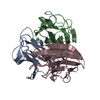
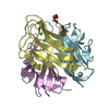
THE STRAIN USED IN THIS WORK HAS NOT BEEN DEPOSITED WITH A SEQUENCE DATABASE, BUT IS EXTREMELY ...THE STRAIN USED IN THIS WORK HAS NOT BEEN DEPOSITED WITH A SEQUENCE DATABASE, BUT IS EXTREMELY CLOSE TO UNIPROT ENTRY Q83WA5. ALL SEQUENCE DIFFERENCES OCCUR IN THE N-TERMINAL PART OF THE MOLECULE, WHICH IS COMPLETELY DISORDERED IN THIS STRUCTURE.

-
Experimental details

-
Experiment

ExperimentMethod: X-RAY DIFFRACTION / Number of used crystals: 1

-
Sample preparation

CrystalDensity Matthews: 2.6 Å3/Da / Density % sol: 52.1 %
Crystal growMethod: vapor diffusion
Details: 20% PEG 4000, 20% ISOPROPANOL, 0.1M NA-CACODYLATE PH = 6.5, 10 MG/ML PROTEIN, VAPOUR DIFFUSION TECHNIQUE

-
Data collection

DiffractionMean temperature: 100 K
Diffraction sourceSource: SYNCHROTRON / Site: ESRF / Beamline: ID14-2 / Wavelength: 0.933
DetectorType: ADSC CCD / Detector: CCD / Date: Jul 30, 2004 / Details: MIRRORS
RadiationMonochromator: CRYSTAL / Protocol: SINGLE WAVELENGTH / Monochromatic (M) / Laue (L): M / Scattering type: x-ray
Radiation wavelengthWavelength: 0.933 Å / Relative weight: 1
ReflectionResolution: 1.36→40 Å / Num. obs: 48836 / % possible obs: 99.3 % / Observed criterion σ(I): 0 / Redundancy: 18.5 % / Rmerge(I) obs: 0.05 / Net I/σ(I): 35.4
Reflection shellResolution: 1.36→1.45 Å / Redundancy: 9.9 % / Rmerge(I) obs: 0.45 / Mean I/σ(I) obs: 4.24 / % possible all: 96

-
Processing

Software
NameVersionClassification
REFMAC5.2.0005refinement
XDSdata reduction
XSCALEdata scaling
autoSHARPphasing
Refinement
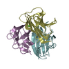
Method to determine structure: SIRAS / Resolution: 1.36→81.65 Å / Cor.coef. Fo:Fc: 0.969 / Cor.coef. Fo:Fc free: 0.969 / SU B: 0.669 / SU ML: 0.027 / Cross valid method: THROUGHOUT / ESU R: 0.042 / ESU R Free: 0.042 / Stereochemistry target values: MAXIMUM LIKELIHOOD
Details: HYDROGENS HAVE BEEN ADDED IN THE RIDING POSITIONS. 79 N-TERMINAL RESIDUES AND 5 C-TERMINAL RESIDUES (HIS-TAG) ARE DISORDERED
RfactorNum. reflection% reflectionSelection details
Rfree0.19 2474 5.1 %RANDOM
Rwork0.179 ---
obs0.179 46407 99.3 %-
Solvent computationIon probe radii: 0.8 Å / Shrinkage radii: 0.8 Å / VDW probe radii: 1.2 Å / Solvent model: MASK
Displacement parametersBiso mean: 17.98 Å2
Baniso -1Baniso -2Baniso -3
1-0.78 Å20.39 Å20 Å2
2--0.78 Å20 Å2
3----1.18 Å2
Refinement stepCycle: LAST / Resolution: 1.36→81.65 Å
ProteinNucleic acidLigandSolventTotal
Num. atoms954 0 5 190 1149
Refine LS restraints
Refine-IDTypeDev idealDev ideal targetNumber
X-RAY DIFFRACTIONr_bond_refined_d0.0080.022993
X-RAY DIFFRACTIONr_bond_other_d
X-RAY DIFFRACTIONr_angle_refined_deg1.161.9911361
X-RAY DIFFRACTIONr_angle_other_deg
X-RAY DIFFRACTIONr_dihedral_angle_1_deg6.3495135
X-RAY DIFFRACTIONr_dihedral_angle_2_deg33.71827.14328
X-RAY DIFFRACTIONr_dihedral_angle_3_deg11.16315156
X-RAY DIFFRACTIONr_dihedral_angle_4_deg
X-RAY DIFFRACTIONr_chiral_restr0.0780.2181
X-RAY DIFFRACTIONr_gen_planes_refined0.0040.02705
X-RAY DIFFRACTIONr_gen_planes_other
X-RAY DIFFRACTIONr_nbd_refined0.1850.2391
X-RAY DIFFRACTIONr_nbd_other
X-RAY DIFFRACTIONr_nbtor_refined0.2990.2710
X-RAY DIFFRACTIONr_nbtor_other
X-RAY DIFFRACTIONr_xyhbond_nbd_refined0.0980.2145
X-RAY DIFFRACTIONr_xyhbond_nbd_other
X-RAY DIFFRACTIONr_metal_ion_refined
X-RAY DIFFRACTIONr_metal_ion_other
X-RAY DIFFRACTIONr_symmetry_vdw_refined0.1840.248
X-RAY DIFFRACTIONr_symmetry_vdw_other
X-RAY DIFFRACTIONr_symmetry_hbond_refined0.1560.221
X-RAY DIFFRACTIONr_symmetry_hbond_other
X-RAY DIFFRACTIONr_symmetry_metal_ion_refined
X-RAY DIFFRACTIONr_symmetry_metal_ion_other
X-RAY DIFFRACTIONr_mcbond_it0.6751.5692
X-RAY DIFFRACTIONr_mcbond_other
X-RAY DIFFRACTIONr_mcangle_it1.12821092
X-RAY DIFFRACTIONr_mcangle_other
X-RAY DIFFRACTIONr_scbond_it1.4073336
X-RAY DIFFRACTIONr_scbond_other
X-RAY DIFFRACTIONr_scangle_it1.9564.5269
X-RAY DIFFRACTIONr_scangle_other
X-RAY DIFFRACTIONr_long_range_B_refined
X-RAY DIFFRACTIONr_long_range_B_other
X-RAY DIFFRACTIONr_rigid_bond_restr
X-RAY DIFFRACTIONr_sphericity_free
X-RAY DIFFRACTIONr_sphericity_bonded
LS refinement shellResolution: 1.36→1.4 Å / Total num. of bins used: 20 /
RfactorNum. reflection
Rfree0.269 168
Rwork0.269 3077

+
About Yorodumi

-
News

-
Feb 9, 2022. New format data for meta-information of EMDB entries

New format data for meta-information of EMDB entries

  • Version 3 of the EMDB header file is now the official format.
  • The previous official version 1.9 will be removed from the archive.

Related info.:EMDB header

External links:wwPDB to switch to version 3 of the EMDB data model

-
Aug 12, 2020. Covid-19 info

Covid-19 info

URL: https://pdbjlvh1.pdbj.org/emnavi/covid19.php

New page: Covid-19 featured information page in EM Navigator.

Related info.:Covid-19 info / Mar 5, 2020. Novel coronavirus structure data

+
Mar 5, 2020. Novel coronavirus structure data

Novel coronavirus structure data

Related info.:Yorodumi Speices / Aug 12, 2020. Covid-19 info

External links:COVID-19 featured content - PDBj / Molecule of the Month (242):Coronavirus Proteases

+
Jan 31, 2019. EMDB accession codes are about to change! (news from PDBe EMDB page)

EMDB accession codes are about to change! (news from PDBe EMDB page)

  • The allocation of 4 digits for EMDB accession codes will soon come to an end. Whilst these codes will remain in use, new EMDB accession codes will include an additional digit and will expand incrementally as the available range of codes is exhausted. The current 4-digit format prefixed with “EMD-” (i.e. EMD-XXXX) will advance to a 5-digit format (i.e. EMD-XXXXX), and so on. It is currently estimated that the 4-digit codes will be depleted around Spring 2019, at which point the 5-digit format will come into force.
  • The EM Navigator/Yorodumi systems omit the EMD- prefix.

Related info.:Q: What is EMD? / ID/Accession-code notation in Yorodumi/EM Navigator

External links:EMDB Accession Codes are Changing Soon! / Contact to PDBj

+
Jul 12, 2017. Major update of PDB

Major update of PDB

  • wwPDB released updated PDB data conforming to the new PDBx/mmCIF dictionary.
  • This is a major update changing the version number from 4 to 5, and with Remediation, in which all the entries are updated.
  • In this update, many items about electron microscopy experimental information are reorganized (e.g. em_software).
  • Now, EM Navigator and Yorodumi are based on the updated data.

External links:wwPDB Remediation / Enriched Model Files Conforming to OneDep Data Standards Now Available in the PDB FTP Archive

-
Yorodumi

Thousand views of thousand structures

  • Yorodumi is a browser for structure data from EMDB, PDB, SASBDB, etc.
  • This page is also the successor to EM Navigator detail page, and also detail information page/front-end page for Omokage search.
  • The word "yorodu" (or yorozu) is an old Japanese word meaning "ten thousand". "mi" (miru) is to see.

Related info.:EMDB / PDB / SASBDB / Comparison of 3 databanks / Yorodumi Search / Aug 31, 2016. New EM Navigator & Yorodumi / Yorodumi Papers / Jmol/JSmol / Function and homology information / Changes in new EM Navigator and Yorodumi

Read more