 Movie
Movie Controller

[English] 日本語
 Yorodumi
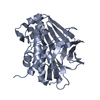
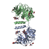
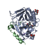
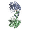
- PDB-1w5r: X-ray crystallographic structure of a C70Q Mycobacterium smegmati... -
+ Open data

- Basic information
| Entry | Database: PDB / ID: 1w5r | ||||||
|---|---|---|---|---|---|---|---|
| Title | X-ray crystallographic structure of a C70Q Mycobacterium smegmatis N- arylamine Acetyltransferase | ||||||
 Components Components | ARYLAMINE N-ACETYLTRANSFERASE | ||||||
 Keywords Keywords | TRANSFERASE / ACYLTRANSFERASE | ||||||
| Function / homology |  Function and homology information Function and homology information | ||||||
| Biological species |  MYCOBACTERIUM SMEGMATIS (bacteria) MYCOBACTERIUM SMEGMATIS (bacteria) | ||||||
| Method |  X-RAY DIFFRACTION / SYNCHROTRON / MOLECULAR REPLACEMENT / Resolution: 1.45 Å X-RAY DIFFRACTION / SYNCHROTRON / MOLECULAR REPLACEMENT / Resolution: 1.45 Å | ||||||
 Authors Authors | Holton, S.J. / Sandy, J. / Rodrigues-Lima, F. / Dupret, J.-M. / Bhakta, S. / Noble, M.E.M. / Sim, E. | ||||||
 Citation Citation | Journal: Biochem.J. / Year: 2005 Title: Investigation of the Catalytic Triad of Arylamine N-Acetyltransferases: Essential Residues Required for Acetyl Transfer to Arylamines. Authors: Sandy, J. / Mushtaq, A. / Holton, S.J. / Schartau, P. / Noble, M.E.M. / Sim, E. | ||||||
| History |
|
- Structure visualization
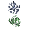
| Structure viewer | Molecule: MolmilJmol/JSmol |
|---|
- Downloads & links
-Download
| PDBx/mmCIF format | 1w5r.cif.gz | 225.5 KB | Display | PDBx/mmCIF format |
|---|---|---|---|---|
| PDB format | pdb1w5r.ent.gz | 182.9 KB | Display | PDB format |
| PDBx/mmJSON format | 1w5r.json.gz | Tree view | PDBx/mmJSON format | |
| Others |  Other downloads Other downloads |
-Validation report
| Arichive directory | https://data.pdbj.org/pub/pdb/validation_reports/w5/1w5rftp://data.pdbj.org/pub/pdb/validation_reports/w5/1w5r | HTTPS FTP |
|---|
-Related structure data
| Related structure data | |
|---|---|
| Similar structure data |


-Links
 PDBj
PDBj- Assembly
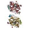
| Deposited unit | 
| ||||||||
|---|---|---|---|---|---|---|---|---|---|
| 1 |
| ||||||||
| Unit cell |
|
-Components
| #1: Protein | Mass: 30591.309 Da / Num. of mol.: 2 / Mutation: YES Source method: isolated from a genetically manipulated source Source: (gene. exp.) MYCOBACTERIUM SMEGMATIS (bacteria) / Production host: #2: Water | ChemComp-HOH / |  Mass: 18.015 Da / Num. of mol.: 398 / Source method: isolated from a natural source / Formula: H2O Mass: 18.015 Da / Num. of mol.: 398 / Source method: isolated from a natural source / Formula: H2OCompound details | ENGINEERED | Sequence details | THE PROTEIN IS PURIFIED USING AN N-TERMINAL HIS-TAG, WHICH IS CLEAVED OFF PRIOR TO CRYSTALLIZATION. ...THE PROTEIN IS PURIFIED USING AN N-TERMINAL HIS-TAG, WHICH IS CLEAVED OFF PRIOR TO CRYSTALLIZ | |
|---|
-Experimental details
-Experiment
| Experiment | Method: X-RAY DIFFRACTION |
|---|
- Sample preparation
| Crystal | Density Matthews: 2.61 Å3/Da / Density % sol: 52.93 % |
|---|
-Data collection
| Diffraction | Mean temperature: 100 K |
|---|---|
| Diffraction source | Source: SYNCHROTRON / Site: ESRF  / Beamline: ID14-2 / Wavelength: 0.934 / Beamline: ID14-2 / Wavelength: 0.934 |
| Detector | Type: ADSC CCD / Detector: CCD |
| Radiation | Protocol: SINGLE WAVELENGTH / Monochromatic (M) / Laue (L): M / Scattering type: x-ray |
| Radiation wavelength | Wavelength: 0.934 Å / Relative weight: 1 |
| Reflection | Resolution: 1.3→31.01 Å / Num. obs: 103538 / % possible obs: 90.5 % / Observed criterion σ(I): 0 / Redundancy: 3.19 % / Rmerge(I) obs: 0.06 / Net I/σ(I): 6.79 |
| Reflection shell | Resolution: 1.45→1.53 Å / Redundancy: 2.72 % / Rmerge(I) obs: 0.41 / Mean I/σ(I) obs: 1.77 / % possible all: 84.7 |
- Processing
| Software | Name: REFMAC / Version: 5.2.0005 24/04/2001 / Classification: refinement | ||||||||||||||||||||||||||||||||||||||||||||||||||||||||||||||||||||||||||||||||||||||||||||||||||||||||||||||||||||||||||||||||||||||||||||||||||||||||||||||||||||||||||||||||||||||
|---|---|---|---|---|---|---|---|---|---|---|---|---|---|---|---|---|---|---|---|---|---|---|---|---|---|---|---|---|---|---|---|---|---|---|---|---|---|---|---|---|---|---|---|---|---|---|---|---|---|---|---|---|---|---|---|---|---|---|---|---|---|---|---|---|---|---|---|---|---|---|---|---|---|---|---|---|---|---|---|---|---|---|---|---|---|---|---|---|---|---|---|---|---|---|---|---|---|---|---|---|---|---|---|---|---|---|---|---|---|---|---|---|---|---|---|---|---|---|---|---|---|---|---|---|---|---|---|---|---|---|---|---|---|---|---|---|---|---|---|---|---|---|---|---|---|---|---|---|---|---|---|---|---|---|---|---|---|---|---|---|---|---|---|---|---|---|---|---|---|---|---|---|---|---|---|---|---|---|---|---|---|---|---|
| Refinement | Method to determine structure: MOLECULAR REPLACEMENT / Resolution: 1.45→79.06 Å / Cor.coef. Fo:Fc: 0.956 / Cor.coef. Fo:Fc free: 0.946 / SU B: 2.401 / SU ML: 0.042 / Cross valid method: THROUGHOUT / ESU R: 0.086 / ESU R Free: 0.073 / Stereochemistry target values: MLF / Details: HYDROGENS HAVE BEEN ADDED IN THE RIDING POSITIONS.
| ||||||||||||||||||||||||||||||||||||||||||||||||||||||||||||||||||||||||||||||||||||||||||||||||||||||||||||||||||||||||||||||||||||||||||||||||||||||||||||||||||||||||||||||||||||||
| Solvent computation | Ion probe radii: 0.8 Å / Shrinkage radii: 0.8 Å / VDW probe radii: 1.2 Å / Solvent model: MASK BULK SOLVENT | ||||||||||||||||||||||||||||||||||||||||||||||||||||||||||||||||||||||||||||||||||||||||||||||||||||||||||||||||||||||||||||||||||||||||||||||||||||||||||||||||||||||||||||||||||||||
| Displacement parameters | Biso mean: 17.95 Å2
| ||||||||||||||||||||||||||||||||||||||||||||||||||||||||||||||||||||||||||||||||||||||||||||||||||||||||||||||||||||||||||||||||||||||||||||||||||||||||||||||||||||||||||||||||||||||
| Refinement step | Cycle: LAST / Resolution: 1.45→79.06 Å
| ||||||||||||||||||||||||||||||||||||||||||||||||||||||||||||||||||||||||||||||||||||||||||||||||||||||||||||||||||||||||||||||||||||||||||||||||||||||||||||||||||||||||||||||||||||||
| Refine LS restraints |
|