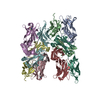
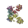
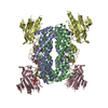
TRANSFERASE / OGT / GLCNAC / NUCLEOPORIN / O-LINKED GLYCOSYLATION / TPR REPEAT / PROTEIN BINDING / SIGNAL TRANSDUCTION
Function / homology
Function and homology information
negative regulation of non-canonical inflammasome complex assembly / protein N-acetylglucosaminyltransferase complex / regulation of insulin receptor signaling pathway / protein O-GlcNAc transferase / protein O-acetylglucosaminyltransferase activity / positive regulation of transcription from RNA polymerase II promoter by glucose / acetylglucosaminyltransferase activity / regulation of Rac protein signal transduction / regulation of necroptotic process / negative regulation of stem cell population maintenance ...negative regulation of non-canonical inflammasome complex assembly / protein N-acetylglucosaminyltransferase complex / regulation of insulin receptor signaling pathway / protein O-GlcNAc transferase / protein O-acetylglucosaminyltransferase activity / positive regulation of transcription from RNA polymerase II promoter by glucose / acetylglucosaminyltransferase activity / regulation of Rac protein signal transduction / regulation of necroptotic process / negative regulation of stem cell population maintenance / protein O-linked glycosylation / NSL complex / regulation of glycolytic process / regulation of gluconeogenesis / RIPK1-mediated regulated necrosis / Formation of WDR5-containing histone-modifying complexes / regulation of synapse assembly / Sin3-type complex / positive regulation of stem cell population maintenance / regulation of neurotransmitter receptor localization to postsynaptic specialization membrane / positive regulation of proteolysis / phosphatidylinositol-3,4,5-trisphosphate binding / hemopoiesis / histone acetyltransferase complex / positive regulation of lipid biosynthetic process / mitophagy / cell projection / response to nutrient / positive regulation of TORC1 signaling / negative regulation of protein ubiquitination / negative regulation of proteasomal ubiquitin-dependent protein catabolic process / negative regulation of cell migration / positive regulation of translation / circadian regulation of gene expression / negative regulation of transforming growth factor beta receptor signaling pathway / cellular response to glucose stimulus / protein processing / chromatin DNA binding / response to insulin / Regulation of necroptotic cell death / mitochondrial membrane / UCH proteinases / positive regulation of cold-induced thermogenesis / HATs acetylate histones / chromatin organization / apoptotic process / regulation of transcription by RNA polymerase II / positive regulation of DNA-templated transcription / glutamatergic synapse / negative regulation of transcription by RNA polymerase II / signal transduction / positive regulation of transcription by RNA polymerase II / protein-containing complex / nucleoplasm / nucleus / plasma membrane / cytosol Similarity search - Function
THE AUTHORS HAVE INDICATED THAT THERE IS EXPERIMENTALEVIDENCE AVAILABLE FROM GEL FILTRATION EXPERIMENTS THATTHIS PROTEIN IS A CONSTITUTIVE DIMER IN SOLUTION, WHICHIS DISCUSSED IN THE JOURNAL REFERENCE ABOVE.
Mass: 18.015 Da / Num. of mol.: 27 / Source method: isolated from a natural source / Formula: H2O
Sequence details
PRO14 AND MET15 IN BOTH CHAINS A AND B ARE DERIVED FROM THE EXPRESSION PLASMID AND THEREFORE DO NOT ...PRO14 AND MET15 IN BOTH CHAINS A AND B ARE DERIVED FROM THE EXPRESSION PLASMID AND THEREFORE DO NOT CORRESPOND TO THE UNIPROT DATABASE ENTRY O15294
-
Experimental details
-
Experiment
Experiment
Method: X-RAY DIFFRACTION / Number of used crystals: 1
In the structure databanks used in Yorodumi, some data are registered as the other names, "COVID-19 virus" and "2019-nCoV". Here are the details of the virus and the list of structure data.
Jan 31, 2019. EMDB accession codes are about to change! (news from PDBe EMDB page)
EMDB accession codes are about to change! (news from PDBe EMDB page)
The allocation of 4 digits for EMDB accession codes will soon come to an end. Whilst these codes will remain in use, new EMDB accession codes will include an additional digit and will expand incrementally as the available range of codes is exhausted. The current 4-digit format prefixed with “EMD-” (i.e. EMD-XXXX) will advance to a 5-digit format (i.e. EMD-XXXXX), and so on. It is currently estimated that the 4-digit codes will be depleted around Spring 2019, at which point the 5-digit format will come into force.
The EM Navigator/Yorodumi systems omit the EMD- prefix.
Related info.:Q: What is EMD? / ID/Accession-code notation in Yorodumi/EM Navigator
Yorodumi is a browser for structure data from EMDB, PDB, SASBDB, etc.
This page is also the successor to EM Navigator detail page, and also detail information page/front-end page for Omokage search.
The word "yorodu" (or yorozu) is an old Japanese word meaning "ten thousand". "mi" (miru) is to see.
Related info.:EMDB / PDB / SASBDB / Comparison of 3 databanks / Yorodumi Search / Aug 31, 2016. New EM Navigator & Yorodumi / Yorodumi Papers / Jmol/JSmol / Function and homology information / Changes in new EM Navigator and Yorodumi
 Movie
Movie Controller

 Yorodumi
 Open data
 Basic information
 Components
 Keywords
 Function and homology information
 HOMO SAPIENS (human)
 X-RAY DIFFRACTION /
  Authors
 Citation
 Structure visualization
 Downloads & links
 Other downloads
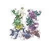


 PDBj
PDBj


 Assembly


 Mass: 40.078 Da / Num. of mol.: 1 / Source method: obtained synthetically / Formula: Ca
 Mass: 18.015 Da / Num. of mol.: 27 / Source method: isolated from a natural source / Formula: H2O
 Sample preparation
 / Beamline: ID14-2 / Wavelength: 0.933
  Processing