 Movie
Movie Controller


+ Open data

- Basic information
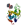
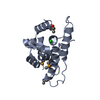
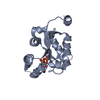
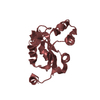
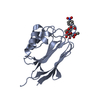
| Entry | Database: PDB / ID: 1vsr | ||||||
|---|---|---|---|---|---|---|---|
| Title | VERY SHORT PATCH REPAIR (VSR) ENDONUCLEASE FROM ESCHERICHIA COLI | ||||||
 Components Components | PROTEIN (VSR ENDONUCLEASE) | ||||||
 Keywords Keywords | HYDROLASE / ENDONUCLEASE / DNA REPAIR / MISMATCH RECOGNITION | ||||||
| Function / homology |  Function and homology information Function and homology informationT/G mismatch-specific endonuclease activity / mismatch repair / Hydrolases; Acting on ester bonds / DNA binding / metal ion binding Similarity search - Function | ||||||
| Biological species |  | ||||||
| Method |  X-RAY DIFFRACTION / SYNCHROTRON / MIR / Resolution: 1.8 Å X-RAY DIFFRACTION / SYNCHROTRON / MIR / Resolution: 1.8 Å | ||||||
 Authors Authors | Tsutakawa, S.E. / Muto, T. / Jingami, H. / Kunishima, N. / Ariyoshi, M. / Kohda, D. / Nakagawa, M. / Morikawa, K. | ||||||
 Citation Citation | Journal: Mol.Cell / Year: 1999 Title: Crystallographic and functional studies of very short patch repair endonuclease. Authors: Tsutakawa, S.E. / Muto, T. / Kawate, T. / Jingami, H. / Kunishima, N. / Ariyoshi, M. / Kohda, D. / Nakagawa, M. / Morikawa, K. | ||||||
| History |
|
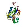
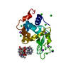
- Structure visualization
| Structure viewer | Molecule: MolmilJmol/JSmol |
|---|
- Downloads & links
-Download
| PDBx/mmCIF format | 1vsr.cif.gz | 40.2 KB | Display | PDBx/mmCIF format |
|---|---|---|---|---|
| PDB format | pdb1vsr.ent.gz | 27.7 KB | Display | PDB format |
| PDBx/mmJSON format | 1vsr.json.gz | Tree view | PDBx/mmJSON format | |
| Others |  Other downloads Other downloads |
-Validation report
| Summary document | 1vsr_validation.pdf.gz | 417.5 KB | Display | wwPDB validaton report |
|---|---|---|---|---|
| Full document | 1vsr_full_validation.pdf.gz | 417.6 KB | Display | |
| Data in XML | 1vsr_validation.xml.gz | 7.9 KB | Display | |
| Data in CIF | 1vsr_validation.cif.gz | 10.2 KB | Display | |
| Arichive directory | https://data.pdbj.org/pub/pdb/validation_reports/vs/1vsrftp://data.pdbj.org/pub/pdb/validation_reports/vs/1vsr | HTTPS FTP |
-Related structure data
| Similar structure data |
|---|

-Links
 PDBj
PDBj


- Assembly
| Deposited unit | 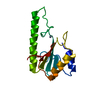
| ||||||||
|---|---|---|---|---|---|---|---|---|---|
| 1 |
| ||||||||
| Unit cell |
|
-Components
| #1: Protein | Mass: 15785.019 Da / Num. of mol.: 1 / Fragment: FRAGMENT Source method: isolated from a genetically manipulated source Source: (gene. exp.) References: UniProt: P09184, Hydrolases; Acting on ester bonds |
|---|---|
| #2: Chemical | ChemComp-ZN /   Mass: 65.409 Da / Num. of mol.: 1 / Source method: obtained synthetically / Formula: Zn Mass: 65.409 Da / Num. of mol.: 1 / Source method: obtained synthetically / Formula: Zn |
| #3: Water | ChemComp-HOH /  Mass: 18.015 Da / Num. of mol.: 63 / Source method: isolated from a natural source / Formula: H2O Mass: 18.015 Da / Num. of mol.: 63 / Source method: isolated from a natural source / Formula: H2O |
-Experimental details
-Experiment
| Experiment | Method: X-RAY DIFFRACTION / Number of used crystals: 1 |
|---|
- Sample preparation
| Crystal | Density Matthews: 2.2 Å3/Da / Density % sol: 44.38 % | ||||||||||||||||||||||||||||||
|---|---|---|---|---|---|---|---|---|---|---|---|---|---|---|---|---|---|---|---|---|---|---|---|---|---|---|---|---|---|---|---|
| Crystal grow | pH: 6.5 Details: CRYSTALLIZATION CONDITIONS: ROOM TEMPERATURE BATCH, 75 MM SODIUM PHOSPHATE PH 6.5, 150 MM NACL, 2 MM DTT, 10% PEG 4K | ||||||||||||||||||||||||||||||
| Crystal grow | *PLUS Temperature: 20 ℃ / Method: batch method | ||||||||||||||||||||||||||||||
| Components of the solutions | *PLUS
|
-Data collection
| Diffraction | Mean temperature: 300 K |
|---|---|
| Diffraction source | Source: SYNCHROTRON / Site: Photon Factory  / Beamline: BL-6B / Wavelength: 1 / Beamline: BL-6B / Wavelength: 1 |
| Detector | Detector: IMAGE PLATE / Date: Nov 17, 1997 / Details: MIRRORS |
| Radiation | Monochromator: SI(111) / Protocol: SINGLE WAVELENGTH / Monochromatic (M) / Laue (L): M / Scattering type: x-ray |
| Radiation wavelength | Wavelength: 1 Å / Relative weight: 1 |
| Reflection | Resolution: 1.8→20 Å / Num. obs: 12869 / % possible obs: 94.8 % / Observed criterion σ(I): 0 / Redundancy: 2.45 % / Rmerge(I) obs: 0.03 / Net I/σ(I): 20.1 |
| Reflection shell | Resolution: 1.8→1.86 Å / Redundancy: 2.3 % / Rmerge(I) obs: 0.177 / % possible all: 91.4 |
| Reflection | *PLUS Rmerge(I) obs: 0.03 |
| Reflection shell | *PLUS % possible obs: 91.4 % |
- Processing
| Software |
| ||||||||||||||||||||
|---|---|---|---|---|---|---|---|---|---|---|---|---|---|---|---|---|---|---|---|---|---|
| Refinement | Method to determine structure: MIR / Resolution: 1.8→20 Å / Cross valid method: THROUGHOUT / σ(F): 0 Details: USED ROUNDS OF XPLOR AND REFMAC FOR REFINEMENT XPLOR-2 SIGMA CUTOFF, 6-1.8 A REFMAC-NO SIGMA CUTOFF, 20-1.8 A, BULK SOLVENT CORRECTION BOND LENGTHS (A) : 0.004 BOND ANGLES (DEGREES) : 2.1
| ||||||||||||||||||||
| Refinement step | Cycle: LAST / Resolution: 1.8→20 Å
| ||||||||||||||||||||
| Software | *PLUS Name: REFMAC / Classification: refinement | ||||||||||||||||||||
| Refinement | *PLUS Highest resolution: 1.8 Å / σ(F): 0 / % reflection Rfree: 5 % / Rfactor obs: 0.2 | ||||||||||||||||||||
| Solvent computation | *PLUS | ||||||||||||||||||||
| Displacement parameters | *PLUS | ||||||||||||||||||||
| Refine LS restraints | *PLUS
|
