 Movie
Movie Controller


+ Open data

- Basic information
| Entry | Database: PDB / ID: 1vr4 | |||||||||
|---|---|---|---|---|---|---|---|---|---|---|
| Title | Crystal Structure of MCSG TArget APC22750 from Bacillus cereus | |||||||||
 Components Components | hypothetical protein APC22750 | |||||||||
 Keywords Keywords | STRUCTURAL GENOMICS / UNKNOWN FUNCTION / Pentamer / Hypothetical Protein / PSI / Protein Structure Initiative / The Midwest Center for Structural Genomics / MCSG | |||||||||
| Function / homology | Uncharacterised protein family UPF0145, YbjQ-like / Putative heavy-metal-binding / UPF0145 domain superfamily / Hypothetical protein apc22750. Chain B / Translation Initiation Factor IF3 / 2-Layer Sandwich / Alpha Beta / UPF0145 protein BC_1012 Function and homology information Function and homology information | |||||||||
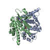
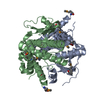
| Biological species |  | |||||||||
| Method |  X-RAY DIFFRACTION / SYNCHROTRON / SAD / Resolution: 2.09 Å X-RAY DIFFRACTION / SYNCHROTRON / SAD / Resolution: 2.09 Å | |||||||||
 Authors Authors | Yang, X. / Brunzelle, J.S. / McNamara, L.K. / Minasov, G. / Shuvalova, L. / Collart, F.R. / Anderson, W.F. / Midwest Center for Structural Genomics (MCSG) | |||||||||
 Citation Citation | Journal: To be Published Title: Crystal Structure of MCSG TArget APC22750 from Bacillus cereus Authors: Brunzelle, J.S. / McNamara, L.K. / Yang, X. / Minasov, G. / Shuvalova, L. / Collart, F.R. / Anderson, W.F. / MCSG | |||||||||
| History |
|
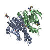
- Structure visualization
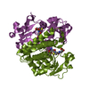
| Structure viewer | Molecule: MolmilJmol/JSmol |
|---|
- Downloads & links
-Download
| PDBx/mmCIF format | 1vr4.cif.gz | 100.2 KB | Display | PDBx/mmCIF format |
|---|---|---|---|---|
| PDB format | pdb1vr4.ent.gz | 79.7 KB | Display | PDB format |
| PDBx/mmJSON format | 1vr4.json.gz | Tree view | PDBx/mmJSON format | |
| Others |  Other downloads Other downloads |
-Validation report
| Arichive directory | https://data.pdbj.org/pub/pdb/validation_reports/vr/1vr4ftp://data.pdbj.org/pub/pdb/validation_reports/vr/1vr4 | HTTPS FTP |
|---|
-Related structure data
| Similar structure data | |
|---|---|
| Other databases |

-Links
 PDBj
PDBj- Assembly
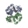
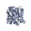
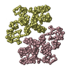
| Deposited unit | 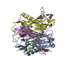
| ||||||||
|---|---|---|---|---|---|---|---|---|---|
| 1 |
| ||||||||
| Unit cell |
|
-Components
| #1: Protein | Mass: 11046.733 Da / Num. of mol.: 5 Source method: isolated from a genetically manipulated source Source: (gene. exp.) #2: Water | ChemComp-HOH / |  Mass: 18.015 Da / Num. of mol.: 151 / Source method: isolated from a natural source / Formula: H2O Mass: 18.015 Da / Num. of mol.: 151 / Source method: isolated from a natural source / Formula: H2O |
|---|
-Experimental details
-Experiment
| Experiment | Method: X-RAY DIFFRACTION / Number of used crystals: 1 |
|---|
- Sample preparation
| Crystal | Density Matthews: 2.2 Å3/Da / Density % sol: 43 % |
|---|---|
| Crystal grow | Temperature: 295 K / Method: vapor diffusion, sitting drop / pH: 8 Details: PEG, pH 8, VAPOR DIFFUSION, SITTING DROP, temperature 295K |
-Data collection
| Diffraction | Mean temperature: 99.8 K |
|---|---|
| Diffraction source | Source: SYNCHROTRON / Site: APS  / Beamline: 32-ID / Wavelength: 0.9792 Å / Beamline: 32-ID / Wavelength: 0.9792 Å |
| Detector | Type: MARRESEARCH / Detector: CCD / Date: Jun 24, 2004 / Details: Mirror |
| Radiation | Monochromator: Diamond 111 / Protocol: SINGLE WAVELENGTH / Monochromatic (M) / Laue (L): M / Scattering type: x-ray |
| Radiation wavelength | Wavelength: 0.9792 Å / Relative weight: 1 |
| Reflection | Resolution: 2.09→30 Å / Num. all: 28306 / Num. obs: 26115 / % possible obs: 92.2 % / Observed criterion σ(F): 0 / Observed criterion σ(I): -3 / Rmerge(I) obs: 0.109 / Net I/σ(I): 12.4 |
| Reflection shell | Resolution: 2.09→2.18 Å / Rmerge(I) obs: 0.27 / Mean I/σ(I) obs: 2.6 / % possible all: 73 |
- Processing
| Software |
| ||||||||||||||||||||||||||||||||||||||||||||||||||||||||||||||||||||||||||||||||||||||||||||||||||||||||||||||||||||||||||||||||||||||||||||||||||||||||||||||||||||||||||
|---|---|---|---|---|---|---|---|---|---|---|---|---|---|---|---|---|---|---|---|---|---|---|---|---|---|---|---|---|---|---|---|---|---|---|---|---|---|---|---|---|---|---|---|---|---|---|---|---|---|---|---|---|---|---|---|---|---|---|---|---|---|---|---|---|---|---|---|---|---|---|---|---|---|---|---|---|---|---|---|---|---|---|---|---|---|---|---|---|---|---|---|---|---|---|---|---|---|---|---|---|---|---|---|---|---|---|---|---|---|---|---|---|---|---|---|---|---|---|---|---|---|---|---|---|---|---|---|---|---|---|---|---|---|---|---|---|---|---|---|---|---|---|---|---|---|---|---|---|---|---|---|---|---|---|---|---|---|---|---|---|---|---|---|---|---|---|---|---|---|---|---|
| Refinement | Method to determine structure: SAD / Resolution: 2.09→25 Å / Cor.coef. Fo:Fc: 0.921 / Cor.coef. Fo:Fc free: 0.861 / SU B: 7.28 / SU ML: 0.192 / TLS residual ADP flag: LIKELY RESIDUAL / Cross valid method: THROUGHOUT / σ(F): 0 / ESU R: 0.335 / ESU R Free: 0.255 / Stereochemistry target values: MAXIMUM LIKELIHOOD
| ||||||||||||||||||||||||||||||||||||||||||||||||||||||||||||||||||||||||||||||||||||||||||||||||||||||||||||||||||||||||||||||||||||||||||||||||||||||||||||||||||||||||||
| Solvent computation | Ion probe radii: 0.8 Å / Shrinkage radii: 0.8 Å / VDW probe radii: 1.4 Å / Solvent model: BABINET MODEL WITH MASK | ||||||||||||||||||||||||||||||||||||||||||||||||||||||||||||||||||||||||||||||||||||||||||||||||||||||||||||||||||||||||||||||||||||||||||||||||||||||||||||||||||||||||||
| Displacement parameters | Biso mean: 11.771 Å2
| ||||||||||||||||||||||||||||||||||||||||||||||||||||||||||||||||||||||||||||||||||||||||||||||||||||||||||||||||||||||||||||||||||||||||||||||||||||||||||||||||||||||||||
| Refinement step | Cycle: LAST / Resolution: 2.09→25 Å
| ||||||||||||||||||||||||||||||||||||||||||||||||||||||||||||||||||||||||||||||||||||||||||||||||||||||||||||||||||||||||||||||||||||||||||||||||||||||||||||||||||||||||||
| Refine LS restraints |
| ||||||||||||||||||||||||||||||||||||||||||||||||||||||||||||||||||||||||||||||||||||||||||||||||||||||||||||||||||||||||||||||||||||||||||||||||||||||||||||||||||||||||||
| LS refinement shell | Resolution: 2.09→2.148 Å / Total num. of bins used: 20 /
| ||||||||||||||||||||||||||||||||||||||||||||||||||||||||||||||||||||||||||||||||||||||||||||||||||||||||||||||||||||||||||||||||||||||||||||||||||||||||||||||||||||||||||
| Refinement TLS params. | Method: refined / Refine-ID: X-RAY DIFFRACTION
| ||||||||||||||||||||||||||||||||||||||||||||||||||||||||||||||||||||||||||||||||||||||||||||||||||||||||||||||||||||||||||||||||||||||||||||||||||||||||||||||||||||||||||
| Refinement TLS group |
|
