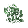
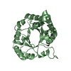
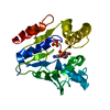
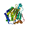
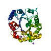
Protein of unknown function UPF0759 / Protein of unknown function DUF72 / UPF0759 superfamily / Protein of unknown function DUF72 / TIM Barrel / Alpha-Beta Barrel / Alpha Beta Similarity search - Domain/homology
Resolution: 2.52→28.52 Å / Num. obs: 13332 / % possible obs: 99.9 % / Redundancy: 6.5 % / Biso Wilson estimate: 53.06 Å2 / Rsym value: 0.18 / Net I/σ(I): 9.1
Reflection shell
Resolution: 2.52→2.66 Å / Redundancy: 6.5 % / Mean I/σ(I) obs: 1.8 / Num. unique all: 1939 / Rsym value: 0.01038 / % possible all: 100
-
Processing
Software
Name
Version
Classification
MOSFLM
datareduction
SCALA
CCP44.2
datascaling
SHARP
phasing
REFMAC
5.2.0005
refinement
CCP4
(SCALA)
datascaling
Refinement
Method to determine structure: MAD / Resolution: 2.52→28.52 Å / Cor.coef. Fo:Fc: 0.92 / Cor.coef. Fo:Fc free: 0.892 / SU B: 21.487 / SU ML: 0.232 / TLS residual ADP flag: LIKELY RESIDUAL / Cross valid method: THROUGHOUT / ESU R: 0.389 / ESU R Free: 0.265 Stereochemistry target values: MAXIMUM LIKELIHOOD WITH PHASES Details: 1) HYDROGENS HAVE BEEN ADDED IN THE RIDING POSITIONS. 2) ELECTRON DENSITY IN TWO REGIONS, RESIDUES 11-19 AND 202-209 IS AMBIGUOUS AND THE STRUCTURE WAS NOT MODELED IN THESE REGIONS. 3) THE ...Details: 1) HYDROGENS HAVE BEEN ADDED IN THE RIDING POSITIONS. 2) ELECTRON DENSITY IN TWO REGIONS, RESIDUES 11-19 AND 202-209 IS AMBIGUOUS AND THE STRUCTURE WAS NOT MODELED IN THESE REGIONS. 3) THE ELECTRON DENSITY FOR THE SIDECHAIN OF CYS 71 INDICATES AN UNIDENTIFIED COVALENT MODIFICATION. 4) THE CRYSTALLIZATION CONTAINS 0.2M ZINC ACETATE; THEREFORE, SEVERAL ZN IONS WERE MODELED INTO ELECTRON DENSITY WITHIN COORDINATION DISTANCE OF POTENTIAL SIDECHAIN LIGANDS (I.E. HISTIDINE AND GLUTAMIC ACID).
Rfactor
Num. reflection
% reflection
Selection details
Rfree
0.25697
657
4.9 %
RANDOM
Rwork
0.22493
-
-
-
obs
0.22652
12658
99.89 %
-
Solvent computation
Ion probe radii: 0.8 Å / Shrinkage radii: 0.8 Å / VDW probe radii: 1.2 Å / Solvent model: BABINET MODEL WITH MASK
Displacement parameters
Biso mean: 38.918 Å2
Baniso -1
Baniso -2
Baniso -3
1-
-2.01 Å2
-1.01 Å2
0 Å2
2-
-
-2.01 Å2
0 Å2
3-
-
-
3.02 Å2
Refinement step
Cycle: LAST / Resolution: 2.52→28.52 Å
Protein
Nucleic acid
Ligand
Solvent
Total
Num. atoms
1998
0
8
37
2043
Refine LS restraints
Refine-ID
Type
Dev ideal
Dev ideal target
Number
X-RAY DIFFRACTION
r_bond_refined_d
0.011
0.022
2061
X-RAY DIFFRACTION
r_angle_refined_deg
1.306
1.933
2799
X-RAY DIFFRACTION
r_dihedral_angle_1_deg
6.34
5
248
X-RAY DIFFRACTION
r_dihedral_angle_2_deg
37.524
24.5
100
X-RAY DIFFRACTION
r_dihedral_angle_3_deg
15.235
15
315
X-RAY DIFFRACTION
r_dihedral_angle_4_deg
23.097
15
7
X-RAY DIFFRACTION
r_chiral_restr
0.085
0.2
303
X-RAY DIFFRACTION
r_gen_planes_refined
0.003
0.02
1594
X-RAY DIFFRACTION
r_nbd_refined
0.197
0.2
929
X-RAY DIFFRACTION
r_xyhbond_nbd_refined
0.123
0.2
92
X-RAY DIFFRACTION
r_symmetry_vdw_refined
0.155
0.2
32
X-RAY DIFFRACTION
r_symmetry_hbond_refined
0.262
0.2
3
X-RAY DIFFRACTION
r_mcbond_it
0.383
1.5
1281
X-RAY DIFFRACTION
r_mcangle_it
0.614
2
2019
X-RAY DIFFRACTION
r_scbond_it
1.246
3
899
X-RAY DIFFRACTION
r_scangle_it
1.653
4.5
780
X-RAY DIFFRACTION
r_nbtor_refined
0.307
0.2
1396
LS refinement shell
Resolution: 2.52→2.586 Å / Total num. of bins used: 20
Rfactor
Num. reflection
% reflection
Rfree
0.416
47
4.81 %
Rwork
0.306
930
-
obs
-
-
100 %
Refinement TLS params.
Method: refined / Refine-ID: X-RAY DIFFRACTION
ID
L11 (°2)
L12 (°2)
L13 (°2)
L22 (°2)
L23 (°2)
L33 (°2)
S11 (Å °)
S12 (Å °)
S13 (Å °)
S21 (Å °)
S22 (Å °)
S23 (Å °)
S31 (Å °)
S32 (Å °)
S33 (Å °)
T11 (Å2)
T12 (Å2)
T13 (Å2)
T22 (Å2)
T23 (Å2)
T33 (Å2)
Origin x (Å)
Origin y (Å)
Origin z (Å)
1
14.1434
-21.8518
5.986
34.5063
-7.4206
7.0195
-1.427
-1.2573
0.1747
3.5434
1.1018
0.8008
-0.9678
-0.4228
0.3252
0.0179
-0.0589
0.0177
0.1276
0.0139
-0.0913
23.8536
25.6633
38.6457
2
3.8112
0.034
-0.2925
5.2299
0.0751
1.8039
0.1273
0.2921
0.1789
-0.397
-0.2142
-0.5659
-0.0664
0.0552
0.0869
-0.0715
0.0304
-0.0163
-0.0981
0.1006
-0.1183
29.7578
32.6765
24.3682
3
9.7998
-3.919
2.0719
7.8959
-1.1067
2.01
0.165
-0.3212
-0.2092
0.0539
-0.1933
0.6452
0.0778
-0.0638
0.0283
-0.0867
-0.0383
-0.0355
-0.0906
-0.0165
-0.1608
14.8819
24.3257
31.863
Refinement TLS group
Refine-ID: X-RAY DIFFRACTION / Selection: ALL / Auth asym-ID: A / Label asym-ID: A
ID
Refine TLS-ID
Auth seq-ID
Label seq-ID
1
1
-3 - 10
9 - 22
2
2
21 - 201
33 - 213
3
3
210 - 265
222 - 277
+
About Yorodumi
-
News
-
Feb 9, 2022. New format data for meta-information of EMDB entries
New format data for meta-information of EMDB entries
Version 3 of the EMDB header file is now the official format.
The previous official version 1.9 will be removed from the archive.
In the structure databanks used in Yorodumi, some data are registered as the other names, "COVID-19 virus" and "2019-nCoV". Here are the details of the virus and the list of structure data.
Jan 31, 2019. EMDB accession codes are about to change! (news from PDBe EMDB page)
EMDB accession codes are about to change! (news from PDBe EMDB page)
The allocation of 4 digits for EMDB accession codes will soon come to an end. Whilst these codes will remain in use, new EMDB accession codes will include an additional digit and will expand incrementally as the available range of codes is exhausted. The current 4-digit format prefixed with “EMD-” (i.e. EMD-XXXX) will advance to a 5-digit format (i.e. EMD-XXXXX), and so on. It is currently estimated that the 4-digit codes will be depleted around Spring 2019, at which point the 5-digit format will come into force.
The EM Navigator/Yorodumi systems omit the EMD- prefix.
Related info.:Q: What is EMD? / ID/Accession-code notation in Yorodumi/EM Navigator
Yorodumi is a browser for structure data from EMDB, PDB, SASBDB, etc.
This page is also the successor to EM Navigator detail page, and also detail information page/front-end page for Omokage search.
The word "yorodu" (or yorozu) is an old Japanese word meaning "ten thousand". "mi" (miru) is to see.
Related info.:EMDB / PDB / SASBDB / Comparison of 3 databanks / Yorodumi Search / Aug 31, 2016. New EM Navigator & Yorodumi / Yorodumi Papers / Jmol/JSmol / Function and homology information / Changes in new EM Navigator and Yorodumi
 Movie
Movie Controller

 Yorodumi
 Open data
 Basic information
 Components
 Keywords
 Function and homology information
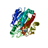
 Enterococcus faecalis (bacteria)
 X-RAY DIFFRACTION /
  Authors
 Citation
 Structure visualization
 Downloads & links
 Other downloads


 PDBj
PDBj

 Assembly

 Mass: 65.409 Da / Num. of mol.: 4 / Source method: obtained synthetically / Formula: Zn
 Mass: 18.015 Da / Num. of mol.: 37 / Source method: isolated from a natural source / Formula: H2O
 Sample preparation
 / Beamline: 8.3.1 / Wavelength: 0.979694, 0.979571, 1.019859
 Processing