 Movie
Movie Controller

[English] 日本語
 Yorodumi
- PDB-1vgg: Crystal Structure of the Conserved Hypothetical Protein TTHA1091 ... -
+ Open data

- Basic information
| Entry | Database: PDB / ID: 1vgg | ||||||
|---|---|---|---|---|---|---|---|
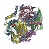
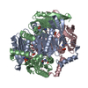
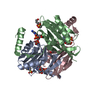
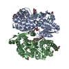
| Title | Crystal Structure of the Conserved Hypothetical Protein TTHA1091 from Thermus Thermophilus HB8 | ||||||
 Components Components | Conserved Hypothetical Protein TT1634 (TTHA1091) | ||||||
 Keywords Keywords | STRUCTURAL GENOMICS / UNKNOWN FUNCTION / Thermus Thermophilus HB8 / Conserved Hypothetical Protein / RIKEN Structural Genomics/Proteomics Initiative / RSGI | ||||||
| Function / homology | Ta1353-like / Adenosine specific kinase / Ta1353-like superfamily / Adenosine specific kinase / hypothetical protein tt1634 / 3-Layer(aba) Sandwich / Alpha Beta / Adenosine monophosphate-protein transferase Function and homology information Function and homology information | ||||||
| Biological species |   Thermus thermophilus HB8 (bacteria) Thermus thermophilus HB8 (bacteria) | ||||||
| Method |  X-RAY DIFFRACTION / SYNCHROTRON / MAD / Resolution: 1.75 Å X-RAY DIFFRACTION / SYNCHROTRON / MAD / Resolution: 1.75 Å | ||||||
 Authors Authors | Satoh, S. / Yao, M. / Kousumi, Y. / Ebihara, A. / Matsumoto, K. / Okamoto, A. / Tanaka, I. / Yokoyama, S. / Kuramitsu, S. / RIKEN Structural Genomics/Proteomics Initiative (RSGI) | ||||||
 Citation Citation | Journal: To be Published Title: Crystal Structure of the Conserved Hypothetical Protein TT1634 from Thermus Thermophilus HB8 Authors: Satoh, S. / Yao, M. / Kousumi, Y. / Ebihara, A. / Matsumoto, K. / Okamoto, A. / Tanaka, I. / Yokoyama, S. / Kuramitsu, S. | ||||||
| History |
|
- Structure visualization
| Structure viewer | Molecule: MolmilJmol/JSmol |
|---|
- Downloads & links
-Download
| PDBx/mmCIF format | 1vgg.cif.gz | 197.6 KB | Display | PDBx/mmCIF format |
|---|---|---|---|---|
| PDB format | pdb1vgg.ent.gz | 158.8 KB | Display | PDB format |
| PDBx/mmJSON format | 1vgg.json.gz | Tree view | PDBx/mmJSON format | |
| Others |  Other downloads Other downloads |
-Validation report
| Summary document | 1vgg_validation.pdf.gz | 462.6 KB | Display | wwPDB validaton report |
|---|---|---|---|---|
| Full document | 1vgg_full_validation.pdf.gz | 469 KB | Display | |
| Data in XML | 1vgg_validation.xml.gz | 37.9 KB | Display | |
| Data in CIF | 1vgg_validation.cif.gz | 54.1 KB | Display | |
| Arichive directory | https://data.pdbj.org/pub/pdb/validation_reports/vg/1vggftp://data.pdbj.org/pub/pdb/validation_reports/vg/1vgg | HTTPS FTP |
-Related structure data
| Similar structure data | |
|---|---|
| Other databases |

-Links
 PDBj
PDBj- Assembly
| Deposited unit | 
| ||||||||
|---|---|---|---|---|---|---|---|---|---|
| 1 |
| ||||||||
| Unit cell |
|
-Components
| #1: Protein | Mass: 17752.438 Da / Num. of mol.: 6 Source method: isolated from a genetically manipulated source Source: (gene. exp.) Thermus thermophilus HB8 (bacteria) / Gene: TTHA1091 / Plasmid: PET11A / Production host: #2: Water | ChemComp-HOH / |  Mass: 18.015 Da / Num. of mol.: 455 / Source method: isolated from a natural source / Formula: H2O Mass: 18.015 Da / Num. of mol.: 455 / Source method: isolated from a natural source / Formula: H2OHas protein modification | Y | |
|---|
-Experimental details
-Experiment
| Experiment | Method: X-RAY DIFFRACTION / Number of used crystals: 1 |
|---|
- Sample preparation
| Crystal | Density Matthews: 2.03 Å3/Da / Density % sol: 38.89 % |
|---|---|
| Crystal grow | Temperature: 293 K / Method: vapor diffusion, sitting drop / pH: 4.6 Details: PEG 200, sodium acetate, sodium chloride, pH 4.6, VAPOR DIFFUSION, SITTING DROP, temperature 293.0K |
-Data collection
| Diffraction | Mean temperature: 100 K | ||||||||||||
|---|---|---|---|---|---|---|---|---|---|---|---|---|---|
| Diffraction source | Source: SYNCHROTRON / Site: SPring-8  / Beamline: BL44B2 / Wavelength: 0.9722, 0.9794, 0.9798 / Beamline: BL44B2 / Wavelength: 0.9722, 0.9794, 0.9798 | ||||||||||||
| Detector | Type: MARRESEARCH / Detector: CCD / Date: Feb 3, 2003 | ||||||||||||
| Radiation | Monochromator: SI 111 / Protocol: MAD / Monochromatic (M) / Laue (L): M / Scattering type: x-ray | ||||||||||||
| Radiation wavelength |
| ||||||||||||
| Reflection | Resolution: 1.75→20 Å / Num. all: 90161 / Num. obs: 90161 / % possible obs: 99 % / Redundancy: 5.3 % / Biso Wilson estimate: 13.3 Å2 / Rmerge(I) obs: 0.057 / Net I/σ(I): 27.9 | ||||||||||||
| Reflection shell | Resolution: 1.75→1.81 Å / Redundancy: 5 % / Rmerge(I) obs: 0.134 / Mean I/σ(I) obs: 9.8 / Num. unique all: 8961 / % possible all: 100 |
- Processing
| Software |
| ||||||||||||||||||||||||||||||||||||||||||||||||||||||||||||||||||||||||||||||||
|---|---|---|---|---|---|---|---|---|---|---|---|---|---|---|---|---|---|---|---|---|---|---|---|---|---|---|---|---|---|---|---|---|---|---|---|---|---|---|---|---|---|---|---|---|---|---|---|---|---|---|---|---|---|---|---|---|---|---|---|---|---|---|---|---|---|---|---|---|---|---|---|---|---|---|---|---|---|---|---|---|---|
| Refinement | Method to determine structure: MAD / Resolution: 1.75→19.93 Å / Rfactor Rfree error: 0.002 / Data cutoff high absF: 2015553.15 / Data cutoff low absF: 0 / Isotropic thermal model: RESTRAINED / Cross valid method: THROUGHOUT / σ(F): 0 / Details: The structure was refined also with Lafire
| ||||||||||||||||||||||||||||||||||||||||||||||||||||||||||||||||||||||||||||||||
| Solvent computation | Solvent model: FLAT MODEL / Bsol: 70.5628 Å2 / ksol: 0.420015 e/Å3 | ||||||||||||||||||||||||||||||||||||||||||||||||||||||||||||||||||||||||||||||||
| Displacement parameters | Biso mean: 16.4 Å2
| ||||||||||||||||||||||||||||||||||||||||||||||||||||||||||||||||||||||||||||||||
| Refine analyze |
| ||||||||||||||||||||||||||||||||||||||||||||||||||||||||||||||||||||||||||||||||
| Refinement step | Cycle: LAST / Resolution: 1.75→19.93 Å
| ||||||||||||||||||||||||||||||||||||||||||||||||||||||||||||||||||||||||||||||||
| Refine LS restraints |
| ||||||||||||||||||||||||||||||||||||||||||||||||||||||||||||||||||||||||||||||||
| LS refinement shell | Resolution: 1.75→1.86 Å / Rfactor Rfree error: 0.006 / Total num. of bins used: 6
| ||||||||||||||||||||||||||||||||||||||||||||||||||||||||||||||||||||||||||||||||
| Xplor file |
|
