 Movie
Movie Controller


+ Open data

- Basic information
| Entry | Database: PDB / ID: 1ve5 | ||||||
|---|---|---|---|---|---|---|---|
| Title | Crystal Structure of T.th. HB8 Threonine deaminase | ||||||
 Components Components | Threonine deaminase | ||||||
 Keywords Keywords | LYASE / threonine deaminase catabolic / RIKEN Structural Genomics/Proteomics Initiative / RSGI / Structural Genomics | ||||||
| Function / homology |  Function and homology information Function and homology informationD-serine biosynthetic process / threonine racemase activity / serine racemase activity / L-serine ammonia-lyase activity / pyridoxal phosphate binding / magnesium ion binding / ATP binding Similarity search - Function | ||||||
| Biological species |   Thermus thermophilus (bacteria) Thermus thermophilus (bacteria) | ||||||
| Method |  X-RAY DIFFRACTION / SYNCHROTRON / MOLECULAR REPLACEMENT / Resolution: 2.15 Å X-RAY DIFFRACTION / SYNCHROTRON / MOLECULAR REPLACEMENT / Resolution: 2.15 Å | ||||||
 Authors Authors | Goto, M. / RIKEN Structural Genomics/Proteomics Initiative (RSGI) | ||||||
 Citation Citation | Journal: To be Published Title: Crystal Structure of T.th. HB8 Threonine deaminase Authors: Goto, M. | ||||||
| History |
|
- Structure visualization
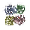
| Structure viewer | Molecule: MolmilJmol/JSmol |
|---|
- Downloads & links
-Download
| PDBx/mmCIF format | 1ve5.cif.gz | 224.8 KB | Display | PDBx/mmCIF format |
|---|---|---|---|---|
| PDB format | pdb1ve5.ent.gz | 181.3 KB | Display | PDB format |
| PDBx/mmJSON format | 1ve5.json.gz | Tree view | PDBx/mmJSON format | |
| Others |  Other downloads Other downloads |
-Validation report
| Arichive directory | https://data.pdbj.org/pub/pdb/validation_reports/ve/1ve5ftp://data.pdbj.org/pub/pdb/validation_reports/ve/1ve5 | HTTPS FTP |
|---|
-Related structure data
| Similar structure data | |
|---|---|
| Other databases |


-Links
 PDBj
PDBj
- Assembly
| Deposited unit | 
| ||||||||
|---|---|---|---|---|---|---|---|---|---|
| 1 |
| ||||||||
| Unit cell |
|
-Components
| #1: Protein | Mass: 32818.926 Da / Num. of mol.: 4 Source method: isolated from a genetically manipulated source Source: (gene. exp.) Thermus thermophilus (bacteria) / Strain: HB8 / Plasmid: pET20b / Production host: #2: Chemical | ChemComp-CA /   Mass: 40.078 Da / Num. of mol.: 4 / Source method: obtained synthetically / Formula: Ca Mass: 40.078 Da / Num. of mol.: 4 / Source method: obtained synthetically / Formula: Ca#3: Chemical | ChemComp-PLP /   Mass: 247.142 Da / Num. of mol.: 4 / Source method: obtained synthetically / Formula: C8H10NO6P Mass: 247.142 Da / Num. of mol.: 4 / Source method: obtained synthetically / Formula: C8H10NO6P#4: Water | ChemComp-HOH / |  Mass: 18.015 Da / Num. of mol.: 256 / Source method: isolated from a natural source / Formula: H2O Mass: 18.015 Da / Num. of mol.: 256 / Source method: isolated from a natural source / Formula: H2O |
|---|
-Experimental details
-Experiment
| Experiment | Method: X-RAY DIFFRACTION / Number of used crystals: 1 |
|---|
- Sample preparation
| Crystal | Density Matthews: 2.46 Å3/Da / Density % sol: 49.97 % |
|---|---|
| Crystal grow | Temperature: 293 K / Method: vapor diffusion, hanging drop / pH: 8.5 Details: PEG4000, MPD, Ca(Ace)2, pH 8.5, VAPOR DIFFUSION, HANGING DROP, temperature 293K |
-Data collection
| Diffraction | Mean temperature: 293 K |
|---|---|
| Diffraction source | Source: SYNCHROTRON / Site: Photon Factory  / Beamline: BL-6A / Wavelength: 1 Å / Beamline: BL-6A / Wavelength: 1 Å |
| Detector | Type: ADSC QUANTUM 4 / Detector: CCD / Date: Jan 31, 2002 |
| Radiation | Protocol: SINGLE WAVELENGTH / Monochromatic (M) / Laue (L): M / Scattering type: x-ray |
| Radiation wavelength | Wavelength: 1 Å / Relative weight: 1 |
| Reflection | Resolution: 2.15→30 Å / Num. all: 67964 / Num. obs: 65660 / % possible obs: 95.7 % / Biso Wilson estimate: 21.4 Å2 |
| Reflection shell | Resolution: 2.15→2.27 Å / % possible all: 95.7 |
- Processing
| Software |
| ||||||||||||||||||||||||||||||||||||
|---|---|---|---|---|---|---|---|---|---|---|---|---|---|---|---|---|---|---|---|---|---|---|---|---|---|---|---|---|---|---|---|---|---|---|---|---|---|
| Refinement | Method to determine structure: MOLECULAR REPLACEMENT / Resolution: 2.15→10 Å / Rfactor Rfree error: 0.003 / Data cutoff high absF: 1479234.77 / Data cutoff low absF: 0 / Isotropic thermal model: RESTRAINED / Cross valid method: THROUGHOUT / σ(F): 0
| ||||||||||||||||||||||||||||||||||||
| Solvent computation | Solvent model: FLAT MODEL / Bsol: 78.4462 Å2 / ksol: 0.424325 e/Å3 | ||||||||||||||||||||||||||||||||||||
| Displacement parameters | Biso mean: 37.9 Å2
| ||||||||||||||||||||||||||||||||||||
| Refine analyze |
| ||||||||||||||||||||||||||||||||||||
| Refinement step | Cycle: LAST / Resolution: 2.15→10 Å
| ||||||||||||||||||||||||||||||||||||
| Refine LS restraints |
| ||||||||||||||||||||||||||||||||||||
| LS refinement shell | Resolution: 2.15→2.28 Å / Rfactor Rfree error: 0.009 / Total num. of bins used: 6
| ||||||||||||||||||||||||||||||||||||
| Xplor file |
|
