 Movie
Movie Controller

[English] 日本語
 Yorodumi
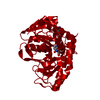
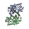
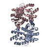
- PDB-1mld: REFINED STRUCTURE OF MITOCHONDRIAL MALATE DEHYDROGENASE FROM PORC... -
+ Open data

- Basic information
| Entry | Database: PDB / ID: 1mld | ||||||
|---|---|---|---|---|---|---|---|
| Title | REFINED STRUCTURE OF MITOCHONDRIAL MALATE DEHYDROGENASE FROM PORCINE HEART AND THE CONSENSUS STRUCTURE FOR DICARBOXYLIC ACID OXIDOREDUCTASES | ||||||
 Components Components | MALATE DEHYDROGENASE | ||||||
 Keywords Keywords | OXIDOREDUCTASE(NAD(A)-CHOH(D)) | ||||||
| Function / homology |  Function and homology information Function and homology information(S)-malate dehydrogenase (NAD+, oxaloacetate-forming) / L-malate dehydrogenase (NAD+) activity / malate metabolic process / tricarboxylic acid cycle / aerobic respiration / protein-folding chaperone binding / mitochondrial matrix / protein homodimerization activity / mitochondrion / cytoplasm Similarity search - Function | ||||||
| Biological species |  | ||||||
| Method |  X-RAY DIFFRACTION / Resolution: 1.83 Å X-RAY DIFFRACTION / Resolution: 1.83 Å | ||||||
 Authors Authors | Gleason, W.B. / Fu, Z. / Birktoft, J.J. / Banaszak, L.J. | ||||||
 Citation Citation | Journal: Biochemistry / Year: 1994 Title: Refined crystal structure of mitochondrial malate dehydrogenase from porcine heart and the consensus structure for dicarboxylic acid oxidoreductases. Authors: Gleason, W.B. / Fu, Z. / Birktoft, J. / Banaszak, L. #1: Journal: Biochemistry / Year: 1993Title: Determinants of Protein Thermostability Observed in the 1.9 Angstrom Crystal Structure of Malate Dehydrogenase from the Thermophilic Bacterium Thermus Flavus Authors: Kelly, C.A. / Nishiyama, M. / Ohnishi, Y. / Beppu, T. / Birktoft, J.J. #2: Journal: Biochemistry / Year: 1989Title: Refined Crystal Structure of Cytoplasmic Malate Dehydrogenase at 2.5 Angstroms Resolution Authors: Birktoft, J.J. / Rhodes, G. / Banaszak, L.J. #3: Journal: Biochem.Soc.Trans. / Year: 1989Title: Comparison of the Molecular Structures of Cytoplasmic and Mitochondrial Malate Dehydrogenases Authors: Birktoft, J.J. / Fu, Z. / Carnahan, G.E. / Rodes, G. / Roderick, S.L. / Banaszak, L.J. #4: Journal: J.Biol.Chem. / Year: 1986Title: The Three Dimensional Structure of Porcine Heart Mitochondrial Malate Dehydrogenase at 3.0 Angstrom Resolution Authors: Roderick, S.L. / Banaszak, L.J. | ||||||
| History |
|
- Structure visualization
| Structure viewer | Molecule: MolmilJmol/JSmol |
|---|
- Downloads & links
-Download
| PDBx/mmCIF format | 1mld.cif.gz | 242 KB | Display | PDBx/mmCIF format |
|---|---|---|---|---|
| PDB format | pdb1mld.ent.gz | 198.8 KB | Display | PDB format |
| PDBx/mmJSON format | 1mld.json.gz | Tree view | PDBx/mmJSON format | |
| Others |  Other downloads Other downloads |
-Validation report
| Arichive directory | https://data.pdbj.org/pub/pdb/validation_reports/ml/1mldftp://data.pdbj.org/pub/pdb/validation_reports/ml/1mld | HTTPS FTP |
|---|
-Related structure data
| Similar structure data |
|---|


-Links
 PDBj
PDBj


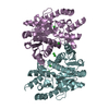
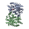
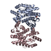
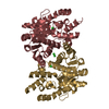
- Assembly
| Deposited unit | 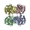
| ||||||||
|---|---|---|---|---|---|---|---|---|---|
| 1 | 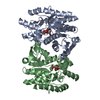
| ||||||||
| 2 | 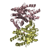
| ||||||||
| 3 |
| ||||||||
| Unit cell |
| ||||||||
| Atom site foot note | 1: CIS PROLINE - PRO A 119 / 2: CIS PROLINE - PRO A 190 / 3: CIS PROLINE - PRO B 119 / 4: CIS PROLINE - PRO B 190 / 5: CIS PROLINE - PRO C 119 / 6: CIS PROLINE - PRO C 190 / 7: CIS PROLINE - PRO D 119 / 8: CIS PROLINE - PRO D 190 |
-Components
| #1: Protein | Mass: 33120.531 Da / Num. of mol.: 4 Source method: isolated from a genetically manipulated source Source: (gene. exp.) References: UniProt: P00346, (S)-malate dehydrogenase (NAD+, oxaloacetate-forming) #2: Chemical | ChemComp-CIT /   Mass: 192.124 Da / Num. of mol.: 4 / Source method: obtained synthetically / Formula: C6H8O7 Mass: 192.124 Da / Num. of mol.: 4 / Source method: obtained synthetically / Formula: C6H8O7#3: Water | ChemComp-HOH / |  Mass: 18.015 Da / Num. of mol.: 521 / Source method: isolated from a natural source / Formula: H2O Mass: 18.015 Da / Num. of mol.: 521 / Source method: isolated from a natural source / Formula: H2O |
|---|
-Experimental details
-Experiment
| Experiment | Method: X-RAY DIFFRACTION |
|---|
- Sample preparation
| Crystal | Density Matthews: 2.59 Å3/Da / Density % sol: 52.43 % | ||||||||||||
|---|---|---|---|---|---|---|---|---|---|---|---|---|---|
| Crystal grow | *PLUS pH: 5.8 / Method: unknownDetails: referred to 'Roderick, S.L.', (1986) J.Biol.Chem., 261, 9461-9464 | ||||||||||||
| Components of the solutions | *PLUS
|
-Data collection
| Radiation | Scattering type: x-ray |
|---|---|
| Radiation wavelength | Relative weight: 1 |
| Reflection | *PLUS Highest resolution: 1.83 Å / Num. obs: 91000 / Rmerge(I) obs: 0.145 |
- Processing
| Software |
| ||||||||||||||||||||||||||||||||||||||||||||||||||||||||||||
|---|---|---|---|---|---|---|---|---|---|---|---|---|---|---|---|---|---|---|---|---|---|---|---|---|---|---|---|---|---|---|---|---|---|---|---|---|---|---|---|---|---|---|---|---|---|---|---|---|---|---|---|---|---|---|---|---|---|---|---|---|---|
| Refinement | Resolution: 1.83→6 Å / σ(F): 0 /
| ||||||||||||||||||||||||||||||||||||||||||||||||||||||||||||
| Refinement step | Cycle: LAST / Resolution: 1.83→6 Å
| ||||||||||||||||||||||||||||||||||||||||||||||||||||||||||||
| Refine LS restraints |
| ||||||||||||||||||||||||||||||||||||||||||||||||||||||||||||
| Software | *PLUS Name: X-PLOR / Classification: refinement | ||||||||||||||||||||||||||||||||||||||||||||||||||||||||||||
| Refinement | *PLUS Rfactor obs: 0.211 / Rfactor Rwork: 0.211 | ||||||||||||||||||||||||||||||||||||||||||||||||||||||||||||
| Solvent computation | *PLUS | ||||||||||||||||||||||||||||||||||||||||||||||||||||||||||||
| Displacement parameters | *PLUS | ||||||||||||||||||||||||||||||||||||||||||||||||||||||||||||
| Refine LS restraints | *PLUS Type: x_angle_d / Dev ideal: 3.2 |
