 Movie
Movie Controller


+ Open data

- Basic information
| Entry | Database: PDB / ID: 1vdr | ||||||
|---|---|---|---|---|---|---|---|
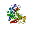
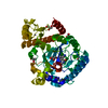
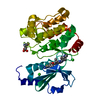
| Title | DIHYDROFOLATE REDUCTASE | ||||||
 Components Components | DIHYDROFOLATE REDUCTASE | ||||||
 Keywords Keywords | OXIDOREDUCTASE / DIHYDROFOLATE REDUCTASE / HALOPHILIC ENZYME | ||||||
| Function / homology |  Function and homology information Function and homology informationdihydrofolate metabolic process / dihydrofolate reductase / dihydrofolate reductase activity / folic acid metabolic process / tetrahydrofolate biosynthetic process / one-carbon metabolic process / NADP binding / cytosol Similarity search - Function | ||||||
| Biological species |  Haloferax volcanii (archaea) Haloferax volcanii (archaea) | ||||||
| Method |  X-RAY DIFFRACTION / MOLECULAR REPLACEMENT / Resolution: 2.55 Å X-RAY DIFFRACTION / MOLECULAR REPLACEMENT / Resolution: 2.55 Å | ||||||
 Authors Authors | Pieper, U. / Herzberg, O. | ||||||
 Citation Citation | Journal: Structure / Year: 1998 Title: Structural features of halophilicity derived from the crystal structure of dihydrofolate reductase from the Dead Sea halophilic archaeon, Haloferax volcanii. Authors: Pieper, U. / Kapadia, G. / Mevarech, M. / Herzberg, O. | ||||||
| History |
|
- Structure visualization
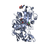
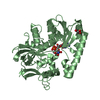
| Structure viewer | Molecule: MolmilJmol/JSmol |
|---|
- Downloads & links
-Download
| PDBx/mmCIF format | 1vdr.cif.gz | 73 KB | Display | PDBx/mmCIF format |
|---|---|---|---|---|
| PDB format | pdb1vdr.ent.gz | 54.9 KB | Display | PDB format |
| PDBx/mmJSON format | 1vdr.json.gz | Tree view | PDBx/mmJSON format | |
| Others |  Other downloads Other downloads |
-Validation report
| Arichive directory | https://data.pdbj.org/pub/pdb/validation_reports/vd/1vdrftp://data.pdbj.org/pub/pdb/validation_reports/vd/1vdr | HTTPS FTP |
|---|
-Related structure data
| Related structure data | 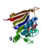 3dfrS 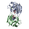 4dfrS  5dfrS 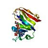 6dfrS  7dfrS  8dfrS S: Starting model for refinement |
|---|---|
| Similar structure data |


-Links
 PDBj
PDBj
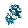
- Assembly
| Deposited unit | 
| ||||||||
|---|---|---|---|---|---|---|---|---|---|
| 1 |
| ||||||||
| Unit cell |
|
-Components
| #1: Protein | Mass: 17997.822 Da / Num. of mol.: 2 / Source method: isolated from a natural source / Details: FORMERLY HALOBACTERIUM VOLCANII / Source: (natural) Haloferax volcanii (archaea) / References: UniProt: P15093, dihydrofolate reductase#2: Chemical |   Mass: 94.971 Da / Num. of mol.: 3 / Source method: obtained synthetically / Formula: PO4 Mass: 94.971 Da / Num. of mol.: 3 / Source method: obtained synthetically / Formula: PO4#3: Water | ChemComp-HOH / |  Mass: 18.015 Da / Num. of mol.: 80 / Source method: isolated from a natural source / Formula: H2O Mass: 18.015 Da / Num. of mol.: 80 / Source method: isolated from a natural source / Formula: H2O |
|---|
-Experimental details
-Experiment
| Experiment | Method: X-RAY DIFFRACTION / Number of used crystals: 1 |
|---|
- Sample preparation
| Crystal | Density Matthews: 2.27 Å3/Da / Density % sol: 45.91 % | ||||||||||||||||||||||||||||||
|---|---|---|---|---|---|---|---|---|---|---|---|---|---|---|---|---|---|---|---|---|---|---|---|---|---|---|---|---|---|---|---|
| Crystal grow | Method: vapor diffusion, hanging drop / pH: 7.9 Details: HANGING DROP VAPOR DIFFUSION. THE RESERVOIR SOLUTION CONTAINED 2.4M PHOSPHATE BUFFER (PH 7.8-8.0), 0.25-0.5% PEG 1000 AND 2MM METHOTREXATE. THE DROPS CONTAINED EQUAL VOLUMES OF RESERVOIR ...Details: HANGING DROP VAPOR DIFFUSION. THE RESERVOIR SOLUTION CONTAINED 2.4M PHOSPHATE BUFFER (PH 7.8-8.0), 0.25-0.5% PEG 1000 AND 2MM METHOTREXATE. THE DROPS CONTAINED EQUAL VOLUMES OF RESERVOIR SOLUTION AND 10 MG/ML PROTEIN SOLUTION IN 0.1M PHOSPHATE BUFFER AT PH7.0., pH 7.9, vapor diffusion - hanging drop PH range: 7.0-8.0 | ||||||||||||||||||||||||||||||
| Crystal grow | *PLUS pH: 7 / Method: vapor diffusion, hanging drop | ||||||||||||||||||||||||||||||
| Components of the solutions | *PLUS
|
-Data collection
| Diffraction | Mean temperature: 295 K |
|---|---|
| Diffraction source | Source: ROTATING ANODE / Type: RIGAKU RUH2R / Wavelength: 1.5418 |
| Detector | Type: SIEMENS / Detector: AREA DETECTOR / Date: Jan 1, 1990 / Details: COLLIMATOR |
| Radiation | Monochromator: GRAPHITE(002) / Monochromatic (M) / Laue (L): M / Scattering type: x-ray |
| Radiation wavelength | Wavelength: 1.5418 Å / Relative weight: 1 |
| Reflection | Resolution: 2.55→50 Å / Num. obs: 10496 / % possible obs: 98 % / Observed criterion σ(I): -3 / Redundancy: 2.8 % / Rmerge(I) obs: 0.073 / Net I/σ(I): 18.2 |
| Reflection shell | Resolution: 2.55→2.71 Å / Redundancy: 1.7 % / Rmerge(I) obs: 0.271 / Mean I/σ(I) obs: 2.4 / % possible all: 89 |
| Reflection shell | *PLUS % possible obs: 88 % |
- Processing
| Software |
| ||||||||||||||||||||||||||||||||||||||||||||||||||||||||||||
|---|---|---|---|---|---|---|---|---|---|---|---|---|---|---|---|---|---|---|---|---|---|---|---|---|---|---|---|---|---|---|---|---|---|---|---|---|---|---|---|---|---|---|---|---|---|---|---|---|---|---|---|---|---|---|---|---|---|---|---|---|---|
| Refinement | Method to determine structure: MOLECULAR REPLACEMENT Starting model: SUPERPOSITION OF 7 MODELS: 4DFR, 5DFR, 6DFR, 7DFR, 8DFR, 3DFR, MODELED HV-DHFR Resolution: 2.55→7 Å / Rfactor Rfree error: 0.01 / Data cutoff high absF: 10000000 / Data cutoff low absF: 0 / Cross valid method: THROUGHOUT / σ(F): 2 Details: THE R-VALUE WITHOUT INCLUDING THE DATA THAT WAS RESERVED FOR THE FREE R CALCULATION THROUGHOUT IS 0.180.
| ||||||||||||||||||||||||||||||||||||||||||||||||||||||||||||
| Displacement parameters | Biso mean: 26 Å2 | ||||||||||||||||||||||||||||||||||||||||||||||||||||||||||||
| Refinement step | Cycle: LAST / Resolution: 2.55→7 Å
| ||||||||||||||||||||||||||||||||||||||||||||||||||||||||||||
| Refine LS restraints |
| ||||||||||||||||||||||||||||||||||||||||||||||||||||||||||||
| Software | *PLUS Name: X-PLOR / Version: 3.1 / Classification: refinement | ||||||||||||||||||||||||||||||||||||||||||||||||||||||||||||
| Refinement | *PLUS Rfactor Rfree: 0.3 | ||||||||||||||||||||||||||||||||||||||||||||||||||||||||||||
| Solvent computation | *PLUS | ||||||||||||||||||||||||||||||||||||||||||||||||||||||||||||
| Displacement parameters | *PLUS |
