 Movie
Movie Controller

[English] 日本語
 Yorodumi
- PDB-1vdm: Crystal structure of purine phosphoribosyltransferase from Pyroco... -
+ Open data

- Basic information
| Entry | Database: PDB / ID: 1vdm | ||||||
|---|---|---|---|---|---|---|---|
| Title | Crystal structure of purine phosphoribosyltransferase from Pyrococcus horikoshii Ot3 | ||||||
 Components Components | purine phosphoribosyltransferase | ||||||
 Keywords Keywords | TRANSFERASE / purine phosphoribosyltransferase / Pyrococcus horikoshii / Structural Genomics / RIKEN Structural Genomics/Proteomics Initiative / RSGI | ||||||
| Function / homology | Rossmann fold - #2020 / Phosphoribosyl transferase domain / Phosphoribosyltransferase-like / Phosphoribosyltransferase domain / glycosyltransferase activity / Rossmann fold / 3-Layer(aba) Sandwich / Alpha Beta / 153aa long hypothetical purine phosphoribosyltransferase Function and homology information Function and homology information | ||||||
| Biological species |   Pyrococcus horikoshii (archaea) Pyrococcus horikoshii (archaea) | ||||||
| Method |  X-RAY DIFFRACTION / SYNCHROTRON / MIR / Resolution: 2.5 Å X-RAY DIFFRACTION / SYNCHROTRON / MIR / Resolution: 2.5 Å | ||||||
 Authors Authors | Sugahara, M. / Kunishima, N. / RIKEN Structural Genomics/Proteomics Initiative (RSGI) | ||||||
 Citation Citation | Journal: To be Published Title: Crystal structure of purine phosphoribosyltransferase from Pyrococcus horikoshii Ot3 Authors: Sugahara, M. / Kunishima, N. | ||||||
| History |
|
- Structure visualization
| Structure viewer | Molecule: MolmilJmol/JSmol |
|---|
- Downloads & links
-Download
| PDBx/mmCIF format | 1vdm.cif.gz | 365.6 KB | Display | PDBx/mmCIF format |
|---|---|---|---|---|
| PDB format | pdb1vdm.ent.gz | 303.7 KB | Display | PDB format |
| PDBx/mmJSON format | 1vdm.json.gz | Tree view | PDBx/mmJSON format | |
| Others |  Other downloads Other downloads |
-Validation report
| Arichive directory | https://data.pdbj.org/pub/pdb/validation_reports/vd/1vdmftp://data.pdbj.org/pub/pdb/validation_reports/vd/1vdm | HTTPS FTP |
|---|
-Related structure data
| Similar structure data | |
|---|---|
| Other databases |

-Links
 PDBj
PDBj
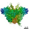
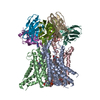
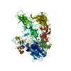
- Assembly
| Deposited unit | 
| ||||||||
|---|---|---|---|---|---|---|---|---|---|
| 1 |
| ||||||||
| Unit cell |
| ||||||||
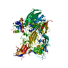
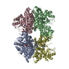
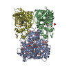
| Details | The biological assembly is a dodecamer in the asymmetric unit. |
-Components
| #1: Protein | Mass: 17601.727 Da / Num. of mol.: 12 Source method: isolated from a genetically manipulated source Source: (gene. exp.) Pyrococcus horikoshii (archaea) / Plasmid: pET11a / Production host:  References: UniProt: O57827, Transferases; Glycosyltransferases; Pentosyltransferases #2: Water | ChemComp-HOH / |  Mass: 18.015 Da / Num. of mol.: 749 / Source method: isolated from a natural source / Formula: H2O Mass: 18.015 Da / Num. of mol.: 749 / Source method: isolated from a natural source / Formula: H2O |
|---|
-Experimental details
-Experiment
| Experiment | Method: X-RAY DIFFRACTION / Number of used crystals: 3 |
|---|
- Sample preparation
| Crystal | Density Matthews: 3.36 Å3/Da / Density % sol: 63.35 % |
|---|---|
| Crystal grow | Temperature: 291 K / Method: microbatch / pH: 4.4 / Details: NaCl, pH 4.4, microbatch, temperature 291K |
-Data collection
| Diffraction |
| ||||||||||||||||||||||||
|---|---|---|---|---|---|---|---|---|---|---|---|---|---|---|---|---|---|---|---|---|---|---|---|---|---|
| Diffraction source |
| ||||||||||||||||||||||||
| Detector |
| ||||||||||||||||||||||||
| Radiation | Protocol: SINGLE WAVELENGTH / Monochromatic (M) / Laue (L): M / Scattering type: x-ray | ||||||||||||||||||||||||
| Radiation wavelength |
| ||||||||||||||||||||||||
| Reflection | Resolution: 2.5→40 Å / Num. all: 99412 / Num. obs: 99412 / % possible obs: 100 % / Observed criterion σ(F): 0 / Observed criterion σ(I): 0 / Redundancy: 7.3 % / Biso Wilson estimate: 50.611 Å2 / Rmerge(I) obs: 0.129 / Rsym value: 0.123 / Net I/σ(I): 8 | ||||||||||||||||||||||||
| Reflection shell | Resolution: 2.5→2.59 Å / Redundancy: 7.4 % / Rmerge(I) obs: 0.785 / Mean I/σ(I) obs: 3.23 / Num. unique all: 9841 / Rsym value: 0.731 / % possible all: 100 |

- Processing
| Software |
| |||||||||||||||||||||||||
|---|---|---|---|---|---|---|---|---|---|---|---|---|---|---|---|---|---|---|---|---|---|---|---|---|---|---|
| Refinement | Method to determine structure: MIR / Resolution: 2.5→39.55 Å / Isotropic thermal model: anisotropic / Cross valid method: THROUGHOUT / σ(F): 0 / Stereochemistry target values: Engh & Huber
| |||||||||||||||||||||||||
| Displacement parameters | Biso mean: 46.4 Å2
| |||||||||||||||||||||||||
| Refine analyze |
| |||||||||||||||||||||||||
| Refinement step | Cycle: LAST / Resolution: 2.5→39.55 Å
| |||||||||||||||||||||||||
| Refine LS restraints |
| |||||||||||||||||||||||||
| LS refinement shell | Resolution: 2.5→2.59 Å / Rfactor Rfree error: 0.014
|
