 Movie
Movie Controller

[English] 日本語
 Yorodumi
- PDB-1v9t: Structure of E. coli cyclophilin B K163T mutant bound to succinyl... -
+ Open data

- Basic information
| Entry | Database: PDB / ID: 1v9t | ||||||
|---|---|---|---|---|---|---|---|
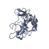
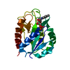
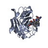
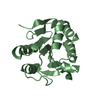
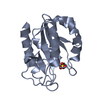
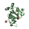
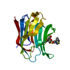
| Title | Structure of E. coli cyclophilin B K163T mutant bound to succinyl-ALA-PRO-ALA-P-nitroanilide | ||||||
 Components Components |
| ||||||
 Keywords Keywords | ISOMERASE/ISOMERASE INHIBITOR / BETA BARREL / ISOMERASE-ISOMERASE INHIBITOR complex | ||||||
| Function / homology |  Function and homology information Function and homology informationprotein peptidyl-prolyl isomerization / peptidylprolyl isomerase / peptidyl-prolyl cis-trans isomerase activity / outer membrane-bounded periplasmic space / protein folding / periplasmic space Similarity search - Function | ||||||
| Biological species |  | ||||||
| Method |  X-RAY DIFFRACTION / SYNCHROTRON / MIR / Resolution: 1.7 Å X-RAY DIFFRACTION / SYNCHROTRON / MIR / Resolution: 1.7 Å | ||||||
 Authors Authors | Konno, M. / Sano, Y. / Okudaira, K. / Kawaguchi, Y. / Yamagishi-Ohmori, Y. / Fushinobu, S. / Matsuzawa, H. | ||||||
 Citation Citation | Journal: Eur.J.Biochem. / Year: 2004 Title: Escherichia coli cyclophilin B binds a highly distorted form of trans-prolyl peptide isomer Authors: Konno, M. / Sano, Y. / Okudaira, K. / Kawaguchi, Y. / Yamagishi-Ohmori, Y. / Fushinobu, S. / Matsuzawa, H. | ||||||
| History |
|
- Structure visualization
| Structure viewer | Molecule: MolmilJmol/JSmol |
|---|
- Downloads & links
-Download
| PDBx/mmCIF format | 1v9t.cif.gz | 78.5 KB | Display | PDBx/mmCIF format |
|---|---|---|---|---|
| PDB format | pdb1v9t.ent.gz | 58.7 KB | Display | PDB format |
| PDBx/mmJSON format | 1v9t.json.gz | Tree view | PDBx/mmJSON format | |
| Others |  Other downloads Other downloads |
-Validation report
| Arichive directory | https://data.pdbj.org/pub/pdb/validation_reports/v9/1v9tftp://data.pdbj.org/pub/pdb/validation_reports/v9/1v9t | HTTPS FTP |
|---|
-Related structure data
| Related structure data |  1j2aC  1vaiC  1lopS C: citing same article ( S: Starting model for refinement |
|---|---|
| Similar structure data |


-Links
 PDBj
PDBj

- Assembly
| Deposited unit | 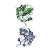
| ||||||||
|---|---|---|---|---|---|---|---|---|---|
| 1 | 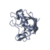
| ||||||||
| 2 | 
| ||||||||
| Unit cell |
|
-Components
| #1: Protein | Mass: 18070.326 Da / Num. of mol.: 2 / Mutation: K163T Source method: isolated from a genetically manipulated source Source: (gene. exp.) References: UniProt: P20752, UniProt: P0AFL3*PLUS, peptidylprolyl isomerase #2: Protein/peptide | ( | 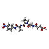  Type: Peptide-like / Class: Inhibitor / Mass: 477.467 Da / Num. of mol.: 1 / Source method: obtained synthetically / Details: Chemically synthesized / References: SUCCINYL-ALA-PRO-ALA-P-NITROANILIDE Type: Peptide-like / Class: Inhibitor / Mass: 477.467 Da / Num. of mol.: 1 / Source method: obtained synthetically / Details: Chemically synthesized / References: SUCCINYL-ALA-PRO-ALA-P-NITROANILIDE#3: Water | ChemComp-HOH / |  Mass: 18.015 Da / Num. of mol.: 181 / Source method: isolated from a natural source / Formula: H2O Mass: 18.015 Da / Num. of mol.: 181 / Source method: isolated from a natural source / Formula: H2O |
|---|
-Experimental details
-Experiment
| Experiment | Method: X-RAY DIFFRACTION / Number of used crystals: 2 |
|---|
- Sample preparation
| Crystal | Density Matthews: 2.71 Å3/Da / Density % sol: 54.18 % |
|---|---|
| Crystal grow | Temperature: 293 K / Method: vapor diffusion, hanging drop / pH: 8 Details: ammonium sulfate, methanol, sodium azide, Tris-HCl, pH 8.0, VAPOR DIFFUSION, HANGING DROP, temperature 293K |
-Data collection
| Diffraction | Mean temperature: 293 K |
|---|---|
| Diffraction source | Source: SYNCHROTRON / Site: Photon Factory  / Beamline: BL-6A / Wavelength: 1 Å / Beamline: BL-6A / Wavelength: 1 Å |
| Detector | Type: WEISSENBERG / Detector: DIFFRACTOMETER / Date: Jan 1, 1999 |
| Radiation | Monochromator: SI / Protocol: SINGLE WAVELENGTH / Monochromatic (M) / Laue (L): M / Scattering type: x-ray |
| Radiation wavelength | Wavelength: 1 Å / Relative weight: 1 |
| Reflection | Resolution: 1.7→80 Å / Num. all: 40355 / Num. obs: 40355 / % possible obs: 92 % / Observed criterion σ(I): 1 / Rmerge(I) obs: 0.09 / Net I/σ(I): 16.7 |
| Reflection shell | Resolution: 1.7→1.76 Å / Rmerge(I) obs: 0.238 / % possible all: 73.3 |
- Processing
| Software |
| ||||||||||||||||||||
|---|---|---|---|---|---|---|---|---|---|---|---|---|---|---|---|---|---|---|---|---|---|
| Refinement | Method to determine structure: MIR Starting model: PDB ENTRY 1LOP Resolution: 1.7→6 Å / σ(F): 2
| ||||||||||||||||||||
| Refinement step | Cycle: LAST / Resolution: 1.7→6 Å
| ||||||||||||||||||||
| Refine LS restraints |
|
